Nature Nanotechnology ( IF 38.3 ) Pub Date : 2023-05-22 , DOI: 10.1038/s41565-023-01401-7 Jiaqi Guo 1 , Fengbin Wang 2, 3, 4 , Yimeng Huang 1 , Hongjian He 1 , Weiyi Tan 1 , Meihui Yi 1 , Edward H Egelman 2 , Bing Xu 1
|
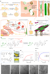
Cell spheroids bridge the discontinuity between in vitro systems and in vivo animal models. However, inducing cell spheroids by nanomaterials remains an inefficient and poorly understood process. Here we use cryogenic electron microscopy to determine the atomic structure of helical nanofibres self-assembled from enzyme-responsive d-peptides and fluorescent imaging to show that the transcytosis of d-peptides induces intercellular nanofibres/gels that potentially interact with fibronectin to enable cell spheroid formation. Specifically, d-phosphopeptides, being protease resistant, undergo endocytosis and endosomal dephosphorylation to generate helical nanofibres. On secretion to the cell surface, these nanofibres form intercellular gels that act as artificial matrices and facilitate the fibrillogenesis of fibronectins to induce cell spheroids. No spheroid formation occurs without endo- or exocytosis, phosphate triggers or shape switching of the peptide assemblies. This study—coupling transcytosis and morphological transformation of peptide assemblies—demonstrates a potential approach for regenerative medicine and tissue engineering.
中文翻译:
通过转胞吞细胞间凝胶化创建细胞球体
细胞球体弥合了体外系统和体内动物模型之间的不连续性。然而,通过纳米材料诱导细胞球体仍然是一个低效且知之甚少的过程。在这里,我们使用低温电子显微镜来确定由酶响应d肽自组装的螺旋纳米纤维的原子结构,并使用荧光成像来表明d肽的转胞吞作用诱导细胞间纳米纤维/凝胶,这些纳米纤维/凝胶可能与纤连蛋白相互作用,从而使细胞球体形成形成。具体而言,具有蛋白酶抗性的d-磷酸肽会经历胞吞作用和内体去磷酸化,从而产生螺旋纳米纤维。在分泌到细胞表面时,这些纳米纤维形成细胞间凝胶,充当人工基质并促进纤连蛋白的原纤维形成,从而诱导细胞球体。如果没有内吞作用或胞吐作用、磷酸盐触发或肽组装体的形状转换,则不会发生球状体形成。这项研究将转胞吞作用和肽组装体的形态转化结合起来,展示了再生医学和组织工程的潜在方法。


 京公网安备 11010802027423号