Surgical Endoscopy ( IF 2.4 ) Pub Date : 2022-09-16 , DOI: 10.1007/s00464-022-09603-1 Yaoping Zhang 1, 2 , Lina Qu 1, 2 , Yani Gou 1, 2 , Jinyong Hao 1, 2 , Yanglin Pan 3 , Xiaojun Huang 1, 2
|
Background
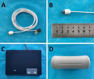
Magnetically controlled capsule endoscopy (MCCE) has recently increasingly been used for gastric examination. However, the image quality and esophageal observation is suboptimal. We developed a novel wired transmission magnetically controlled capsule endoscopy (WT-MCCE) system and evaluated its feasibility through in vitro and in vivo experiments.
Methods
A plastic stomach model and a pathological upper gastrointestinal model were used to evaluate the performance of WT-MCCE in vitro experiments. Twice of examination in the two in vitro models by WT-MCCE were performed by 5 endoscopists who were experienced in performing wireless capsule endoscopy. The examination of traditional gastroscopy (Olympus, GIF-HQ290) in the pathological upper gastrointestinal model was set as the control. In vivo experiments were performed in a live canine model by 3 endoscopists, in which WT-MCCE was inserted with the assistance of gastroscopy. Measurements included maneuverability, examination time, visualization of gastric mucosa, image quality and diagnostic accuracy.
Results
WT-MCCE showed good performance in both in vitro and in vivo experiments with excellent visualization of mucosa (75–100%). The mean operation time is 17.6 ± 2.7 min, 22.3 ± 1.9 min and 29.3 ± 3.4 min in three models, respectively. In pathological upper gastrointestinal model, all lesions, including esophageal varices, one polyp, one foreign body, two gastric ulcers and one duodenal ulcer, were detected by both WT-MCCE and traditional gastroscopy by all endoscopists. For the observation of esophagus and stomach in the canine model, WT-MCCE also showed excellent maneuverability and good image quality.
Conclusions
The novel WT-MCCE system performed well in evaluating upper gastrointestinal landmarks and lesions in two in vitro models, and showed good performance in a canine model. WT-MCCE may be potentially useful for diagnosis of esophageal and gastric diseases.
Graphical abstract
中文翻译:
新型有线传输磁控胶囊内窥镜系统用于上消化道检查的体外和体内评价
背景
磁控胶囊内窥镜(MCCE)最近越来越多地用于胃检查。然而,图像质量和食管观察并不理想。我们开发了一种新型有线传输磁控胶囊内窥镜(WT-MCCE)系统,并通过体外和体内实验评估其可行性。
方法
采用塑料胃模型和病理上消化道模型来评估WT-MCCE体外实验的性能。 WT-MCCE 对两个体外模型进行了两次检查,由 5 名具有无线胶囊内窥镜经验的内窥镜医师进行。以传统胃镜检查(Olympus,GIF-HQ290)在上消化道病理模型中的检查作为对照。 3 名内窥镜医师在活体犬模型上进行了体内实验,在胃镜检查的帮助下将 WT-MCCE 插入其中。测量包括可操作性、检查时间、胃粘膜可视化、图像质量和诊断准确性。
结果
WT-MCCE 在体外和体内实验中均表现出良好的性能,具有出色的粘膜可视化效果 (75–100%)。三种模型的平均操作时间分别为 17.6 ± 2.7 分钟、22.3 ± 1.9 分钟和 29.3 ± 3.4 分钟。在病理上消化道模型中,所有内镜医师均通过WT-MCCE和传统胃镜检查发现所有病变,包括食管静脉曲张、1例息肉、1例异物、2例胃溃疡和1例十二指肠溃疡。对于犬模型中食道和胃的观察,WT-MCCE也表现出了出色的可操作性和良好的图像质量。
结论
新型WT-MCCE系统在两个体外模型中评估上消化道标志和病变方面表现良好,并且在犬模型中表现出良好的性能。 WT-MCCE 可能对食管和胃疾病的诊断有用。


 京公网安备 11010802027423号