Acta Neuropathologica ( IF 9.3 ) Pub Date : 2022-08-26 , DOI: 10.1007/s00401-022-02476-7 Sara A M Holec 1 , Jisoo Lee 2 , Abby Oehler 2 , Felicia K Ooi 2 , Daniel A Mordes 2, 3 , Steven H Olson 2, 4, 5 , Stanley B Prusiner 2, 4, 6 , Amanda L Woerman 1, 2, 4
|
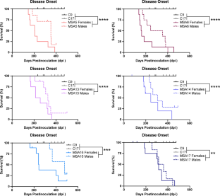
In multiple system atrophy (MSA), the protein α-synuclein misfolds into a prion conformation that self-templates and causes progressive neurodegeneration. While many point mutations in the α-synuclein gene, SNCA, have been identified as the cause of heritable Parkinson’s disease (PD), none have been identified as causing MSA. To examine whether MSA prions can transmit disease to mice expressing wild-type (WT) human α-synuclein, we inoculated transgenic (Tg) mice denoted TgM20+/− with brain homogenates prepared from six different deceased MSA patients. All six samples transmitted CNS disease to the mice, with an average incubation period of ~ 280 days. Interestingly, TgM20+/− female mice developed disease > 60 days earlier than their male counterparts. Brains from terminal mice contained phosphorylated α-synuclein throughout the hindbrain, consistent with the distribution of α-synuclein inclusions in MSA patients. In addition, using our α-syn–YFP cell lines, we detected α-synuclein prions in brain homogenates prepared from terminal mice that retained MSA strain properties. To our knowledge, the studies described here are the first to show that MSA prions transmit neurological disease to mice expressing WT SNCA and that the rate of transmission is sex dependent. By comparison, TgM20+/− mice inoculated with WT preformed fibrils (PFFs) developed severe neurological disease in ~ 210 days and exhibited robust α-synuclein neuropathology in both limbic regions and the hindbrain. Brain homogenates from these animals exhibited biological activities that are distinct from those found in MSA-inoculated mice when tested in the α-syn–YFP cell lines. Differences between brains from MSA-inoculated and WT PFF-inoculated mice potentially argue that α-synuclein prions from MSA patients are distinct from the PFF inocula and that PFFs do not replicate MSA strain biology.
中文翻译:
多系统萎缩朊病毒将神经系统疾病传播给表达野生型人类α-突触核蛋白的小鼠
在多系统萎缩 (MSA) 中,蛋白质 α-突触核蛋白错误折叠成朊病毒构象,自我模板化并导致进行性神经变性。虽然 α-突触核蛋白基因SNCA中的许多点突变已被确定为遗传性帕金森病 (PD) 的病因,但尚未发现任何点突变会导致 MSA。为了检查 MSA 朊病毒是否可以将疾病传播给表达野生型 (WT) 人类 α-突触核蛋白的小鼠,我们用从六名不同的已故 MSA 患者制备的脑匀浆接种了表示为 TgM20 +/-的转基因 (Tg) 小鼠。所有六个样本均将中枢神经系统疾病传播给小鼠,平均潜伏期约为 280 天。有趣的是,TgM20 +/-雌性小鼠比雄性小鼠早 60 天发病。临终小鼠的大脑在整个后脑中含有磷酸化的 α-突触核蛋白,与 MSA 患者中 α-突触核蛋白内含物的分布一致。此外,使用我们的 α-syn-YFP 细胞系,我们在保留 MSA 菌株特性的终末小鼠制备的脑匀浆中检测到 α-突触核蛋白朊病毒。据我们所知,这里描述的研究首次表明 MSA 朊病毒会将神经系统疾病传播给表达 WT SNCA 的小鼠,并且传播率取决于性别。相比之下,接种 WT 预制原纤维 (PFF) 的 TgM20 +/-小鼠在约 210 天内出现严重的神经系统疾病,并在边缘区域和后脑表现出强烈的 α-突触核蛋白神经病理学。在 α-syn-YFP 细胞系中进行测试时,这些动物的脑匀浆表现出与接种 MSA 的小鼠不同的生物活性。MSA 接种小鼠和 WT PFF 接种小鼠大脑之间的差异可能表明,MSA 患者的 α-突触核蛋白朊病毒与 PFF 接种物不同,并且 PFF 不能复制 MSA 菌株生物学。


 京公网安备 11010802027423号