Acta Neuropathologica ( IF 9.3 ) Pub Date : 2022-08-18 , DOI: 10.1007/s00401-022-02463-y Regina J Faubel 1 , Veronica S Santos Canellas 1 , Jenna Gaesser 2 , Nancy H Beluk 3 , Tim N Feinstein 1 , Yong Wang 4 , Maya Yankova 5 , Kalyani B Karunakaran 6 , Stephen M King 5 , Madhavi K Ganapathiraju 7 , Cecilia W Lo 1
|
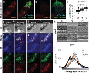
A carpet of ependymal motile cilia lines the brain ventricular system, forming a network of flow channels and barriers that pattern cerebrospinal fluid (CSF) flow at the surface. This CSF transport system is evolutionary conserved, but its physiological function remains unknown. Here we investigated its potential role in epilepsy with studies focused on CDKL5 deficiency disorder (CDD), a neurodevelopmental disorder with early-onset epilepsy refractory to seizure medications and the most common cause of infant epilepsy. CDKL5 is a highly conserved X-linked gene suggesting its function in regulating cilia length and motion in the green alga Chlamydomonas might have implication in the etiology of CDD. Examination of the structure and function of airway motile cilia revealed both the CDD patients and the Cdkl5 knockout mice exhibit cilia lengthening and abnormal cilia motion. Similar defects were observed for brain ventricular cilia in the Cdkl5 knockout mice. Mapping ependymal cilia generated flow in the ventral third ventricle (v3V), a brain region with important physiological functions showed altered patterning of flow. Tracing of cilia-mediated inflow into v3V with fluorescent dye revealed the appearance of a flow barrier at the inlet of v3V in Cdkl5 knockout mice. Analysis of mice with a mutation in another epilepsy-associated kinase, Yes1, showed the same disturbance of cilia motion and flow patterning. The flow barrier was also observed in the Foxj1± and FOXJ1CreERT:Cdkl5y/fl mice, confirming the contribution of ventricular cilia to the flow disturbances. Importantly, mice exhibiting altered cilia-driven flow also showed increased susceptibility to anesthesia-induced seizure-like activity. The cilia-driven flow disturbance arises from altered cilia beating orientation with the disrupted polarity of the cilia anchoring rootlet meshwork. Together these findings indicate motile cilia disturbances have an essential role in CDD-associated seizures and beyond, suggesting cilia regulating kinases may be a therapeutic target for medication-resistant epilepsy.
中文翻译:
血流阻塞扰乱了癫痫脑中由纤毛驱动的液体运输
室管膜运动纤毛地毯排列在脑室系统中,形成流动通道和屏障网络,在表面形成脑脊液 (CSF) 流动模式。这种 CSF 运输系统在进化上是保守的,但其生理功能仍然未知。在这里,我们调查了它在癫痫中的潜在作用,研究重点是 CDKL5 缺乏症 (CDD),这是一种神经发育障碍,伴有早发性癫痫,癫痫药物难以治愈,是婴儿癫痫的最常见原因。CDKL5是一种高度保守的 X 连锁基因,表明其在绿藻衣藻中调节纤毛长度和运动的功能可能与 CDD 的病因学有关。对气道活动纤毛的结构和功能的检查表明,CDD 患者和Cdkl5基因敲除小鼠均表现出纤毛延长和异常纤毛运动。在Cdkl5敲除小鼠的脑室纤毛中观察到类似的缺陷。绘制室管膜纤毛在腹侧第三脑室 (v3V) 中产生的流量,这是一个具有重要生理功能的大脑区域,显示流量模式发生了改变。用荧光染料追踪纤毛介导的流入 v3V 揭示了在Cdkl5敲除小鼠中 v3V 入口处出现流动屏障。对另一种癫痫相关激酶 Yes1 发生突变的小鼠的分析表明,纤毛运动和流动模式存在相同的紊乱。流动障碍也被观察到Foxj1 ±和FOXJ1Cre ERT :Cdkl5 y/fl小鼠,确认心室纤毛对血流障碍的贡献。重要的是,表现出改变的纤毛驱动流动的小鼠也表现出对麻醉诱导的癫痫样活动的易感性增加。纤毛驱动的流动干扰是由改变的纤毛跳动方向和纤毛锚定细根网状结构的极性破坏引起的。这些发现共同表明活动纤毛紊乱在 CDD 相关癫痫发作及其他发作中起着重要作用,表明纤毛调节激酶可能是药物耐药性癫痫的治疗靶点。


 京公网安备 11010802027423号