European Journal of Nuclear Medicine and Molecular Imaging ( IF 8.6 ) Pub Date : 2022-07-05 , DOI: 10.1007/s00259-022-05893-8 Wanqi Chen 1, 2 , Lei Liu 1, 2 , Yinghe Li 1, 2 , Shatong Li 1, 2 , Zhijian Li 1, 2 , Weiguang Zhang 1, 2 , Xu Zhang 1, 2 , Runze Wu 3 , Debin Hu 3 , Hongyan Sun 3 , Yun Zhou 3 , Wei Fan 1, 2 , Yumo Zhao 1, 2 , Yizhuo Zhang 1, 4 , Yingying Hu 1, 2
|
Purpose
To explore the impact of a true half dose of [18F]-FDG on image quality in pediatric oncological patients undergoing total-body PET/CT and investigate short acquisition times with half-dose injected activity.
Methods
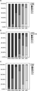
One hundred pediatric oncological patients who underwent total-body PET/CT using the uEXPLORER scanner after receiving a true half dose of [18F]-FDG (1.85 MBq/kg) were retrospectively enrolled. The PET images were first reconstructed using complete 600-s data and then split into 300-s, 180-s, 60-s, 40-s, and 20-s duration groups (G600 to G20). The subjective analysis was performed using 5-point Likert scales. Objective quantitative metrics included the maximum standard uptake value (SUVmax), SUVmean, standard deviation (SD), signal-to-noise ratio (SNR), and SNRnorm of the background. The variabilities in lesion SUVmean, SUVmax, and tumor-to-background ratio (TBR) were also calculated.
Results
The overall image quality scores in the G600, G300, G180, and G60 groups were 4.9 ± 0.2, 4.9 ± 0.3, 4.4 ± 0.5, and 3.5 ± 0.5 points, respectively. All the lesions identified in the half-dose images were localized in the G60 images, while 56% of the lesions could be clearly identified in the G20 images. With reduced acquisition time, the SUVmax and SD of the backgrounds were gradually increased, while the TBR values showed no statistically significant differences among the groups (all p > 0.1). Using the half-dose images as a reference, the variability in the lesion SUVmax gradually increased from the G180 to G20 images, while the lesion SUVmean remained stable across all age groups. SNRnorm was highly negatively correlated with age.
Conclusion
Total-body PET/CT with a half dose of [18F]-FDG (1.85 MBq/kg, estimated whole-body effective dose: 1.76–2.57 mSv) achieved good performance in pediatric patients, with sufficient image quality and good lesion conspicuity. Sufficient image quality and lesion conspicuity could be maintained at a fast scanning time of 60 s with half-dose activity.
中文翻译:
使用半剂量 [18F]-FDG 的全身 PET/CT 评估儿科恶性肿瘤
目的
探讨真正半剂量 [ 18 F]-FDG 对接受全身 PET/CT 的儿科肿瘤患者图像质量的影响,并研究半剂量注射活动的短采集时间。
方法
100 名接受真正半剂量 [ 18 F]-FDG (1.85 MBq/kg)后使用 uEXPLORER 扫描仪接受全身 PET/CT 的儿科肿瘤患者被回顾性纳入。PET 图像首先使用完整的 600 秒数据重建,然后分成 300 秒、180 秒、60 秒、40 秒和 20 秒持续时间组(G600 到 G20)。使用 5 点李克特量表进行主观分析。客观定量指标包括最大标准摄取值 (SUV max )、SUV平均值、标准偏差 (SD)、信噪比 (SNR) 和背景的 SNR范数。病灶SUV mean , SUV max的变异,还计算了肿瘤背景比(TBR)。
结果
G600、G300、G180 和 G60 组的总体图像质量得分分别为 4.9 ± 0.2、4.9 ± 0.3、4.4 ± 0.5 和 3.5 ± 0.5 分。半剂量图像中识别出的所有病灶在 G60 图像中均定位,而在 G20 图像中可以清楚地识别出 56% 的病灶。随着采集时间的减少,背景的 SUV最大值和 SD 逐渐增加,而 TBR 值在各组之间没有统计学差异(所有p > 0.1)。使用半剂量图像作为参考,病变 SUV最大值的变异性从 G180 到 G20 图像逐渐增加,而病变 SUV平均值在所有年龄组中保持稳定。信噪比范数与年龄高度负相关。
结论
半剂量[ 18 F]-FDG(1.85 MBq/kg,估计全身有效剂量:1.76–2.57 mSv)的全身PET/CT在儿科患者中取得了良好的效果,具有足够的图像质量和良好的病灶显着性. 在 60 秒的快速扫描时间和半剂量活动下,可以保持足够的图像质量和病变的显着性。


 京公网安备 11010802027423号