Iranian Journal of Science and Technology, Transactions A: Science ( IF 1.4 ) Pub Date : 2022-06-13 , DOI: 10.1007/s40995-022-01307-4 Seyed Mohammad Taghi Razavi Tousi , Masoomeh Sharifi , Maryam Naseroleslami , Yaser Azizi , Nahid Aboutaleb
|
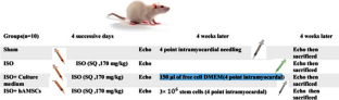
Heart failure (HF) is a leading cause of death that has remained incurable. Recently, stem cell therapy has emerged as a promising tool in cardiac regenerative medicine. Human amniotic membrane-derived mesenchymal stem cells (hAMSCs) with unique characteristics can be used in HF treatment. Here, we aimed to examine the effects of hAMSCs transplantation on cardiac fibrosis in a rat model of ISO-induced HF. Forty male Wistar rats were divided into four groups: sham, isoproterenol-induced HF ((Iso)-)ISO, ISO + culture medium, and ISO + hAMSCs. HF was induced by subcutaneous injection of isoproterenol 170 mg/kg/d in 4 consecutive days. Four weeks later, in ISO + hAMSCs, 3 × 10⁶ hAMSCs were injected into the myocardium, whereas the ISO + culture medium was only injected by cell culture medium. Finally, cardiac functions and hemodynamic parameters were measured. Immunohistochemistry (IHC), Western blot, and histological assessment were performed to evaluate myocardial fibrosis and detect vascular endothelial growth factor (VEGF) collagen type I and III expression level. HF model caused a decrease in ejection fraction (EF) and fraction shortening, whereas both were increased after hAMSCs transplantation. IHC and Western blot and Western blot analyses confirmed that hAMSCs could attenuate fibrosis, reduce collagen I and III depositions, and increase VEGF expression. Intramyocardial transplantation of hAMSCs improves cardiac functions and myocardial structure caused by HF. A rise in VEGF expression presents hAMSCs as a compatible source of stem cell therapy for HF.
中文翻译:
源自人羊膜的间充质干细胞可增加 VEGF 并减轻心力衰竭大鼠的纤维化
心力衰竭 (HF) 是导致死亡的主要原因,仍然无法治愈。最近,干细胞疗法已成为心脏再生医学中一种很有前途的工具。具有独特特性的人羊膜间充质干细胞(hAMSCs)可用于HF治疗。在这里,我们旨在检查 hAMSCs 移植对 ISO 诱导的 HF 大鼠模型中心脏纤维化的影响。将 40 只雄性 Wistar 大鼠分为四组:假手术组、异丙肾上腺素诱导的 HF ((Iso)-)ISO、ISO + 培养基和 ISO + hAMSCs。连续 4 天皮下注射异丙肾上腺素 170 mg/kg/d 诱发 HF。4 周后,在 ISO + hAMSCs 中,将 3 × 10⁶ 个 hAMSCs 注射到心肌中,而 ISO + 培养基仅通过细胞培养基注射。最后,测量心脏功能和血流动力学参数。进行免疫组织化学 (IHC)、蛋白质印迹和组织学评估以评估心肌纤维化并检测血管内皮生长因子 (VEGF) I 型和 III 型胶原蛋白的表达水平。HF 模型导致射血分数 (EF) 降低和分数缩短,而 hAMSCs 移植后两者均增加。IHC 和蛋白质印迹以及蛋白质印迹分析证实,hAMSCs 可以减轻纤维化,减少胶原蛋白 I 和 III 的沉积,并增加 VEGF 的表达。hAMSCs 的心肌内移植可改善由 HF 引起的心脏功能和心肌结构。VEGF 表达的升高使 hAMSCs 成为 HF 干细胞治疗的兼容来源。进行组织学评估以评估心肌纤维化并检测血管内皮生长因子(VEGF)I型和III型胶原的表达水平。HF 模型导致射血分数 (EF) 降低和分数缩短,而 hAMSCs 移植后两者均增加。IHC 和蛋白质印迹以及蛋白质印迹分析证实,hAMSCs 可以减轻纤维化,减少胶原蛋白 I 和 III 的沉积,并增加 VEGF 的表达。hAMSCs 的心肌内移植可改善由 HF 引起的心脏功能和心肌结构。VEGF 表达的升高使 hAMSCs 成为 HF 干细胞治疗的兼容来源。进行组织学评估以评估心肌纤维化并检测血管内皮生长因子(VEGF)I型和III型胶原的表达水平。HF 模型导致射血分数 (EF) 降低和分数缩短,而 hAMSCs 移植后两者均增加。IHC 和蛋白质印迹以及蛋白质印迹分析证实,hAMSCs 可以减轻纤维化,减少胶原蛋白 I 和 III 的沉积,并增加 VEGF 的表达。hAMSCs 的心肌内移植可改善由 HF 引起的心脏功能和心肌结构。VEGF 表达的升高使 hAMSCs 成为 HF 干细胞治疗的兼容来源。HF 模型导致射血分数 (EF) 降低和分数缩短,而 hAMSCs 移植后两者均增加。IHC 和蛋白质印迹以及蛋白质印迹分析证实,hAMSCs 可以减轻纤维化,减少胶原蛋白 I 和 III 的沉积,并增加 VEGF 的表达。hAMSCs 的心肌内移植可改善由 HF 引起的心脏功能和心肌结构。VEGF 表达的升高使 hAMSCs 成为 HF 干细胞治疗的兼容来源。HF 模型导致射血分数 (EF) 降低和分数缩短,而 hAMSCs 移植后两者均增加。IHC 和蛋白质印迹以及蛋白质印迹分析证实,hAMSCs 可以减轻纤维化,减少胶原蛋白 I 和 III 的沉积,并增加 VEGF 的表达。hAMSCs 的心肌内移植可改善由 HF 引起的心脏功能和心肌结构。VEGF 表达的升高使 hAMSCs 成为 HF 干细胞治疗的兼容来源。


 京公网安备 11010802027423号