Interdisciplinary Sciences: Computational Life Sciences ( IF 3.9 ) Pub Date : 2021-06-17 , DOI: 10.1007/s12539-021-00452-5 Xiaoli Zhou 1 , Chaowei Tang 1 , Pan Huang 1 , Francesco Mercaldo 2 , Antonella Santone 2 , Yanqing Shao 3
|
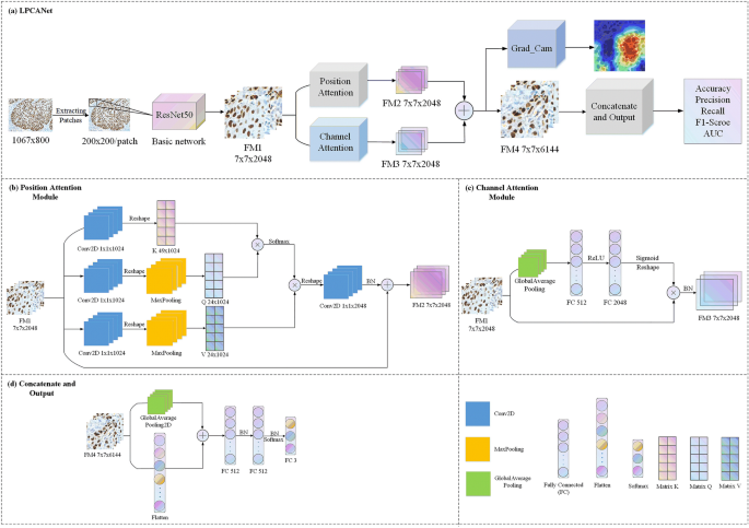
Laryngeal cancer is one of the most common malignant tumors in otolaryngology, and histopathological image analysis is the gold standard for the diagnosis of laryngeal cancer. However, pathologists have high subjectivity in their diagnoses, which makes it easy to miss diagnoses and misdiagnose. In addition, according to a literature search, there is currently no computer-aided diagnosis (CAD) algorithm that has been applied to the classification of histopathological images of laryngeal cancer. Convolutional neural networks (CNNs) are widely used in various other cancer classification tasks. However, the potential global and channel relationships of images may be ignored, which will affect the feature representation ability. Simultaneously, due to the lack of interpretability, the results are often difficult to accept by pathologists. we propose a laryngeal cancer classification network (LPCANet) based on a CNN and attention mechanisms. First, the original histopathological images are sequentially cropped into patches. Then, the patches are input into the basic ResNet50 to extract the local features. Then, a position attention module and a channel attention module are added in parallel to capture the spatial dependency and the channel dependency, respectively. The two modules produce the fusion feature map to enhance the feature representation and improve network classification performance. Moreover, the fusion feature map is extracted and visually analyzed by the grad-weighted class activation map (Grad_CAM) to provide a certain interpretability for the final results. The three-class classification performance of LPCANet is better than those of the five state-of-the-art classifiers (VGG16, ResNet50, InceptionV3, Xception and DenseNet121) on the two original resolutions (534 * 400 and 1067 * 800). On the 534 * 400 data, LPCANet achieved 73.18% accuracy, 74.04% precision, 73.15% recall, 72.9% F1-score, and 0.8826 AUC. On the 1067 * 800 data, LPCANet achieved 83.15% accuracy, 83.5% precision, 83.1% recall, 83.1% F1-score, and 0.9487 AUC. The results show that LPCANet enhances the feature representation by capturing the global and channel relationships and achieves better classification performance. In addition, the visual analysis of Grad_CAM makes the results interpretable, which makes it easier for the results to be accepted by pathologists and allows the method to become a second tool for auxiliary diagnosis.
Graphic Abstract
中文翻译:
LPCANet:使用具有位置注意和通道注意机制的 CNN 对喉癌组织病理学图像进行分类
喉癌是耳鼻喉科最常见的恶性肿瘤之一,组织病理学图像分析是喉癌诊断的金标准。然而,病理学家在诊断上具有较高的主观性,容易漏诊误诊。此外,根据文献检索,目前还没有计算机辅助诊断(CAD)算法应用于喉癌组织病理学图像的分类。卷积神经网络 (CNN) 广泛用于各种其他癌症分类任务。然而,可能会忽略图像潜在的全局和通道关系,这会影响特征表示能力。同时,由于缺乏可解释性,结果往往难以被病理学家接受。我们提出了一个基于 CNN 和注意力机制的喉癌分类网络 (LPCANet)。首先,原始的组织病理学图像被依次裁剪成补丁。然后,将补丁输入到基本的 ResNet50 中以提取局部特征。然后,并行添加位置注意力模块和通道注意力模块,分别捕获空间依赖和通道依赖。这两个模块产生融合特征图以增强特征表示并提高网络分类性能。此外,融合特征图通过梯度加权类激活图(Grad_CAM)进行提取和可视化分析,为最终结果提供一定的可解释性。在两个原始分辨率(534*400和1067*800)上,LPCANet的三类分类性能优于五个最先进的分类器(VGG16、ResNet50、InceptionV3、Xception和DenseNet121)。在534*400的数据上,LPCANet达到了73.18%的准确率、74.04%的准确率、73.15%的召回率、72.9%的F1-score和0.8826的AUC。在 1067 * 800 数据上,LPCANet 实现了 83.15% 的准确率、83.5% 的准确率、83.1% 的召回率、83.1% 的 F1-score 和 0.9487 AUC。结果表明,LPCANet通过捕捉全局和通道关系增强了特征表示,取得了更好的分类性能。此外,Grad_CAM 的可视化分析使结果具有可解释性,这使得结果更容易被病理学家接受,并使该方法成为辅助诊断的第二工具。


 京公网安备 11010802027423号