Analytical and Bioanalytical Chemistry ( IF 3.8 ) Pub Date : 2020-09-02 , DOI: 10.1007/s00216-020-02858-4 Vitalii Serdiuk 1, 2, 3 , Kristen L Shogren 1 , Tetiana Kovalenko 3 , Bakhtiyor Rasulev 2 , Michael Yaszemski 1 , Avudaiappan Maran 1 , Andriy Voronov 2
|
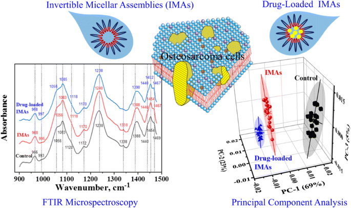
Fourier transform infrared (FTIR) microspectroscopy provides a biochemical fingerprint of the cells. In this study, chemical changes in 143B osteosarcoma cells were investigated using FTIR analysis of cancer cells after their treatment with polymeric invertible micellar assemblies (IMAs) and curcumin-loaded IMAs and compared with untreated osteosarcoma cells. A comprehensive principal component analysis (PCA) was applied to analyze the FTIR results and confirm noticeable changes in cell surface chemical structures in the fingerprint regions of 1480–900 cm−1. The performed clustering shows visible differences for all investigated groups of cancer cells. It is demonstrated that a combination of FTIR microspectroscopy with PCA can be an efficient approach in determining interactions of osteosarcoma cells and drug-loaded polymer micellar assemblies.
中文翻译:
FTIR显微技术检测大分子倒置诱导的骨肉瘤细胞结构变化。
傅里叶变换红外(FTIR)显微光谱提供了细胞的生化指纹。在这项研究中,使用聚合物可逆胶束组件(IMA)和姜黄素负载的IMA处理癌细胞后,使用FTIR分析癌细胞,研究了143B骨肉瘤细胞的化学变化,并与未处理的骨肉瘤细胞进行了比较。应用综合主成分分析(PCA)来分析FTIR结果并确认1480–900 cm -1指纹区域中细胞表面化学结构的显着变化。对于所有研究过的癌细胞组,执行的聚类显示可见的差异。结果表明,FTIR光谱技术与PCA的组合可以成为确定骨肉瘤细胞与载药聚合物胶束组件相互作用的有效方法。


 京公网安备 11010802027423号