当前位置:
X-MOL 学术
›
Nat. Biomed. Eng.
›
论文详情
Our official English website, www.x-mol.net, welcomes your
feedback! (Note: you will need to create a separate account there.)
High-resolution optoacoustic imaging of tissue responses to vascular-targeted therapies.
Nature Biomedical Engineering ( IF 26.8 ) Pub Date : 2020-03-12 , DOI: 10.1038/s41551-020-0527-8 Katja Haedicke 1 , Lilach Agemy 2 , Murad Omar 3, 4 , Andrei Berezhnoi 3, 4 , Sheryl Roberts 5 , Camila Longo-Machado 5 , Magdalena Skubal 1 , Karan Nagar 6 , Hsiao-Ting Hsu 5 , Kwanghee Kim 6 , Thomas Reiner 5, 7 , Jonathan Coleman 6, 8 , Vasilis Ntziachristos 3, 4 , Avigdor Scherz 2 , Jan Grimm 1, 5, 7, 9
Nature Biomedical Engineering ( IF 26.8 ) Pub Date : 2020-03-12 , DOI: 10.1038/s41551-020-0527-8 Katja Haedicke 1 , Lilach Agemy 2 , Murad Omar 3, 4 , Andrei Berezhnoi 3, 4 , Sheryl Roberts 5 , Camila Longo-Machado 5 , Magdalena Skubal 1 , Karan Nagar 6 , Hsiao-Ting Hsu 5 , Kwanghee Kim 6 , Thomas Reiner 5, 7 , Jonathan Coleman 6, 8 , Vasilis Ntziachristos 3, 4 , Avigdor Scherz 2 , Jan Grimm 1, 5, 7, 9
Affiliation
|
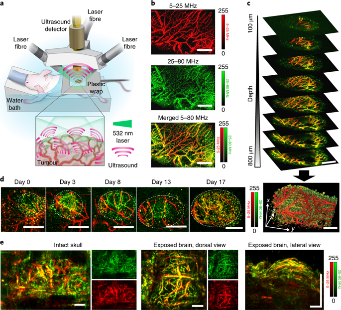
The monitoring of vascular-targeted therapies using magnetic resonance imaging, computed tomography or ultrasound is limited by their insufficient spatial resolution. Here, by taking advantage of the intrinsic optical properties of haemoglobin, we show that raster-scanning optoacoustic mesoscopy (RSOM) provides high-resolution images of the tumour vasculature and of the surrounding tissue, and that the detection of a wide range of ultrasound bandwidths enables the distinction of vessels of differing size, providing detailed insights into the vascular responses to vascular-targeted therapy. Using RSOM to examine the responses to vascular-targeted photodynamic therapy in mice with subcutaneous xenografts, we observed a substantial and immediate occlusion of the tumour vessels followed by haemorrhage within the tissue and the eventual collapse of the entire vasculature. Using dual-wavelength RSOM, which distinguishes oxyhaemoglobin from deoxyhaemoglobin, we observed an increase in oxygenation of the entire tumour volume immediately after the application of the therapy, and a second wave of oxygen reperfusion approximately 24 h thereafter. We also show that RSOM enables the quantification of differences in neoangiogenesis that predict treatment efficacy.
中文翻译:

高分辨率光声成像的组织对血管靶向疗法的反应。
使用磁共振成像,计算机断层扫描或超声对血管靶向疗法的监测受到空间分辨率不足的限制。在这里,通过利用血红蛋白的固有光学特性,我们显示了光栅扫描光声镜(RSOM)提供了肿瘤脉管系统和周围组织的高分辨率图像,以及宽范围的超声带宽检测能够区分不同大小的血管,从而提供对血管靶向治疗的血管反应的详细见解。使用RSOM来检查皮下异种移植小鼠对血管靶向光动力疗法的反应,我们观察到肿瘤血管被实质性的立即阻塞,随后组织内出血,最终整个血管系统塌陷。使用区分氧合血红蛋白和脱氧血红蛋白的双波长RSOM,我们观察到在应用该疗法后立即整个肿瘤体积的氧合增加,此后大约24小时进行第二次氧再灌注。我们还表明,RSOM能够量化预测治疗功效的新血管生成中的差异。
更新日期:2020-04-24
中文翻译:
高分辨率光声成像的组织对血管靶向疗法的反应。
使用磁共振成像,计算机断层扫描或超声对血管靶向疗法的监测受到空间分辨率不足的限制。在这里,通过利用血红蛋白的固有光学特性,我们显示了光栅扫描光声镜(RSOM)提供了肿瘤脉管系统和周围组织的高分辨率图像,以及宽范围的超声带宽检测能够区分不同大小的血管,从而提供对血管靶向治疗的血管反应的详细见解。使用RSOM来检查皮下异种移植小鼠对血管靶向光动力疗法的反应,我们观察到肿瘤血管被实质性的立即阻塞,随后组织内出血,最终整个血管系统塌陷。使用区分氧合血红蛋白和脱氧血红蛋白的双波长RSOM,我们观察到在应用该疗法后立即整个肿瘤体积的氧合增加,此后大约24小时进行第二次氧再灌注。我们还表明,RSOM能够量化预测治疗功效的新血管生成中的差异。


 京公网安备 11010802027423号