当前位置:
X-MOL 学术
›
Nat. Rev. Cardiol.
›
论文详情
Our official English website, www.x-mol.net, welcomes your
feedback! (Note: you will need to create a separate account there.)
Diagnostic imaging of cardiac amyloidosis.
Nature Reviews Cardiology ( IF 41.7 ) Pub Date : 2020-02-10 , DOI: 10.1038/s41569-020-0334-7 Ana Martinez-Naharro 1 , A John Baksi 2, 3 , Philip N Hawkins 1 , Marianna Fontana 1
Nature Reviews Cardiology ( IF 41.7 ) Pub Date : 2020-02-10 , DOI: 10.1038/s41569-020-0334-7 Ana Martinez-Naharro 1 , A John Baksi 2, 3 , Philip N Hawkins 1 , Marianna Fontana 1
Affiliation
|
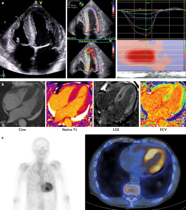
Systemic amyloidosis encompasses a debilitating, under-diagnosed but increasingly recognized group of disorders characterized by the extracellular deposition of misfolded proteins in one or more organs. Cardiac amyloid deposition leads to an infiltrative or restrictive cardiomyopathy and is the major contributor to poor prognosis in patients with systemic amyloidosis. In total, >30 proteins can form amyloid fibrils, but the two main types of amyloid that can infiltrate the heart are monoclonal immunoglobulin light-chain amyloid and transthyretin amyloid. Cardiac amyloidosis can be acquired in older individuals or inherited from birth. Given the nonspecific symptoms of these disorders, a high index of suspicion is paramount in making the correct diagnosis, which can involve the use of non-invasive imaging methods such as echocardiography, bone scintigraphy and cardiovascular MRI. In the past decade, the use of cardiovascular MRI with tissue characterization and bone scintigraphy to diagnose cardiac amyloidosis has revolutionized our understanding of the disease, leading to changes in patient care. However, a need remains for improved awareness and expertise, and greater clinical suspicion, because the initial clues provided by electrocardiography and echocardiography might not be typical. With specific treatments now available, timely diagnosis of cardiac amyloidosis is more important than ever. In this Review, we discuss the current and novel approaches for the diagnostic imaging of cardiac amyloidosis.
中文翻译:

心脏淀粉样变性的诊断影像。
系统性淀粉样变性病包括一组虚弱的,诊断不足的,但得到越来越多认识的疾病,其特征在于错误折叠的蛋白质在一个或多个器官中的细胞外沉积。心脏淀粉样蛋白沉积会导致浸润性或限制性心肌病,并且是导致系统性淀粉样变性病患者预后不良的主要原因。总共,> 30种蛋白质可以形成淀粉样蛋白原纤维,但是可以渗入心脏的两种主要类型的淀粉样蛋白是单克隆免疫球蛋白轻链淀粉样蛋白和运甲状腺素蛋白淀粉样蛋白。心脏淀粉样变性可以在年龄较大的个体中获得或从出生时遗传。鉴于这些疾病的非特异性症状,在做出正确诊断时,高度怀疑是至关重要的,这可能涉及使用非侵入性成像方法,例如超声心动图,骨闪烁显像和心血管MRI。在过去的十年中,将心血管MRI与组织特征和骨闪烁显像技术一起用于诊断心脏淀粉样变性病,彻底改变了我们对这种疾病的理解,从而改变了患者的护理方式。然而,由于心电图和超声心动图提供的最初线索可能并不典型,因此仍然需要提高认识和专业知识,并增加临床怀疑。现在有了特定的治疗方法,及时诊断心脏淀粉样变性比以往任何时候都更为重要。在这篇评论中,我们讨论了心脏淀粉样变性病诊断成像的当前和新颖方法。结合组织特征和骨闪烁显像技术使用心血管MRI诊断心脏淀粉样变性,彻底改变了我们对这种疾病的认识,从而改变了患者的护理方式。然而,由于心电图和超声心动图提供的最初线索可能并不典型,因此仍然需要提高认识和专业知识,并增加临床怀疑。现在有了特定的治疗方法,及时诊断心脏淀粉样变性比以往任何时候都更为重要。在这篇综述中,我们讨论了心脏淀粉样变性病诊断成像的当前和新颖方法。结合组织特征和骨闪烁显像技术使用心血管MRI诊断心脏淀粉样变性,彻底改变了我们对这种疾病的认识,从而改变了患者的护理方式。然而,由于心电图和超声心动图提供的最初线索可能并不典型,因此仍然需要提高认识和专业知识,并增加临床怀疑。现在有了特定的治疗方法,及时诊断心脏淀粉样变性比以往任何时候都更为重要。在这篇评论中,我们讨论了心脏淀粉样变性病诊断成像的当前和新颖方法。因为由心电图和超声心动图提供的最初线索可能并不典型。现在有了特定的治疗方法,及时诊断心脏淀粉样变性比以往任何时候都更为重要。在这篇评论中,我们讨论了心脏淀粉样变性病诊断成像的当前和新颖方法。因为由心电图和超声心动图提供的最初线索可能并不典型。现在有了特定的治疗方法,及时诊断心脏淀粉样变性比以往任何时候都更为重要。在这篇评论中,我们讨论了心脏淀粉样变性病诊断成像的当前和新颖方法。
更新日期:2020-02-10
中文翻译:
心脏淀粉样变性的诊断影像。
系统性淀粉样变性病包括一组虚弱的,诊断不足的,但得到越来越多认识的疾病,其特征在于错误折叠的蛋白质在一个或多个器官中的细胞外沉积。心脏淀粉样蛋白沉积会导致浸润性或限制性心肌病,并且是导致系统性淀粉样变性病患者预后不良的主要原因。总共,> 30种蛋白质可以形成淀粉样蛋白原纤维,但是可以渗入心脏的两种主要类型的淀粉样蛋白是单克隆免疫球蛋白轻链淀粉样蛋白和运甲状腺素蛋白淀粉样蛋白。心脏淀粉样变性可以在年龄较大的个体中获得或从出生时遗传。鉴于这些疾病的非特异性症状,在做出正确诊断时,高度怀疑是至关重要的,这可能涉及使用非侵入性成像方法,例如超声心动图,骨闪烁显像和心血管MRI。在过去的十年中,将心血管MRI与组织特征和骨闪烁显像技术一起用于诊断心脏淀粉样变性病,彻底改变了我们对这种疾病的理解,从而改变了患者的护理方式。然而,由于心电图和超声心动图提供的最初线索可能并不典型,因此仍然需要提高认识和专业知识,并增加临床怀疑。现在有了特定的治疗方法,及时诊断心脏淀粉样变性比以往任何时候都更为重要。在这篇评论中,我们讨论了心脏淀粉样变性病诊断成像的当前和新颖方法。结合组织特征和骨闪烁显像技术使用心血管MRI诊断心脏淀粉样变性,彻底改变了我们对这种疾病的认识,从而改变了患者的护理方式。然而,由于心电图和超声心动图提供的最初线索可能并不典型,因此仍然需要提高认识和专业知识,并增加临床怀疑。现在有了特定的治疗方法,及时诊断心脏淀粉样变性比以往任何时候都更为重要。在这篇综述中,我们讨论了心脏淀粉样变性病诊断成像的当前和新颖方法。结合组织特征和骨闪烁显像技术使用心血管MRI诊断心脏淀粉样变性,彻底改变了我们对这种疾病的认识,从而改变了患者的护理方式。然而,由于心电图和超声心动图提供的最初线索可能并不典型,因此仍然需要提高认识和专业知识,并增加临床怀疑。现在有了特定的治疗方法,及时诊断心脏淀粉样变性比以往任何时候都更为重要。在这篇评论中,我们讨论了心脏淀粉样变性病诊断成像的当前和新颖方法。因为由心电图和超声心动图提供的最初线索可能并不典型。现在有了特定的治疗方法,及时诊断心脏淀粉样变性比以往任何时候都更为重要。在这篇评论中,我们讨论了心脏淀粉样变性病诊断成像的当前和新颖方法。因为由心电图和超声心动图提供的最初线索可能并不典型。现在有了特定的治疗方法,及时诊断心脏淀粉样变性比以往任何时候都更为重要。在这篇评论中,我们讨论了心脏淀粉样变性病诊断成像的当前和新颖方法。


 京公网安备 11010802027423号