npj Aging ( IF 4.1 ) Pub Date : 2017-01-25 , DOI: 10.1038/s41514-017-0002-2 Teru Kamogashira , Ken Hayashi , Chisato Fujimoto , Shinichi Iwasaki , Tatsuya Yamasoba
|
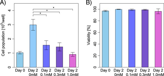
We aimed at determining the mitochondrial function in premature senescence model of auditory cells. Short exposure to H2O2 (1 h, 0.1 mM) induced premature cellular senescence in House Ear Institute-Organ of Corti 1 auditory cells. The transmission electron microscopy analysis revealed that damaged mitochondria and autophagosomes containing dense organelles appeared in the auditory cells after short exposure to H2O2. The branch and junction parameters of the skeletonized image of the mitochondria were found to decrease significantly in H2O2-treated cells. A branched reticulum of tubules was poorly formed, featuring coexistence of numerous tiny clusters along with few relatively large entities in the H2O2-treated cells. In terms of bioenergetics, H2O2-treatment led to the dose-dependent decrease in mitochondrial membrane potential in the auditory cells. The fragmented mitochondria (fusion < fission) were in a low potential. In addition, the potential of hyperfused mitochondria (fusion > fission) was slightly lower than the control cells. The short-time exposure of live auditory cells to H2O2 damaged the mitochondrial respiratory capacity without any effect on the baseline ATP production rates. The vulnerability of the mitochondrial membrane potential to the uncoupling reagent was increased after H2O2 treatment. Our findings indicated that the mitochondrial dysfunction due to the decline in the O2 consumption rate should be the first event of premature senescence process in the auditory cells, resulting in the imbalance of mitochondrial fusion/fission and the collapse of the mitochondrial network.
中文翻译:
在衰老诱导应激下在听觉细胞中观察到功能和形态受损的线粒体
我们旨在确定听觉细胞过早衰老模型中的线粒体功能。在House Ear Institute-Corti 1听觉细胞器官中,短暂暴露于H 2 O 2(1 h,0.1 mM)会导致细胞过早衰老。透射电镜分析表明,短时间暴露于H 2 O 2后,听觉细胞中出现了线粒体和含有致密的细胞器的自噬体。发现H 2 O 2中线粒体骨架化图像的分支和连接参数显着降低处理的细胞。在H 2 O 2处理的细胞中,小管的分支网状结构形成得很差,其特征是许多微小簇与少数相对较大的实体共存。就生物能学而言,H 2 O 2处理导致听觉细胞线粒体膜电位的剂量依赖性降低。破碎的线粒体(融合<裂变)处于低电位。另外,线粒体过度融合的潜力(融合>裂变)略低于对照细胞。活的听觉细胞短时间暴露于H 2 O 2破坏了线粒体的呼吸能力,而对基线ATP的产生速率没有任何影响。H 2 O 2处理后,线粒体膜电位对解偶联剂的脆弱性增加。我们的研究结果表明,由于O 2消耗率下降而引起的线粒体功能障碍应该是听觉细胞过早衰老的首个事件,从而导致线粒体融合/裂变的失衡和线粒体网络的崩溃。


 京公网安备 11010802027423号