International Journal of Oral Science ( IF 10.8 ) Pub Date : 2018-05-17 , DOI: 10.1038/s41368-018-0017-y Alexander Juerchott , Thorsten Pfefferle , Christa Flechtenmacher , Johannes Mente , Martin Bendszus , Sabine Heiland , Tim Hilgenfeld
|
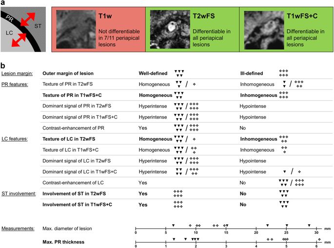
The purpose of this pilot study was to evaluate whether periapical granulomas can be differentiated from periapical cysts in vivo by using dental magnetic resonance imaging (MRI). Prior to apicoectomy, 11 patients with radiographically confirmed periapical lesions underwent dental MRI, including fat-saturated T2-weighted (T2wFS) images, non-contrast-enhanced T1-weighted images with and without fat saturation (T1w/T1wFS), and contrast-enhanced fat-saturated T1-weighted (T1wFS+C) images. Two independent observers performed structured image analysis of MRI datasets twice. A total of 15 diagnostic MRI criteria were evaluated, and histopathological results (6 granulomas and 5 cysts) were compared with MRI characteristics. Statistical analysis was performed using intraclass correlation coefficient (ICC), Cohen’s kappa (κ), Mann–Whitney U-test and Fisher’s exact test. Lesion identification and consecutive structured image analysis was possible on T2wFS and T1wFS+C MRI images. A high reproducibility was shown for MRI measurements of the maximum lesion diameter (intraobserver ICC = 0.996/0.998; interobserver ICC = 0.997), for the “peripheral rim” thickness (intraobserver ICC = 0.988/0.984; interobserver ICC = 0.970), and for all non-quantitative MRI criteria (intraobserver-κ = 0.990/0.995; interobserver-κ = 0.988). In accordance with histopathological results, six MRI criteria allowed for a clear differentiation between cysts and granulomas: (1) outer margin of lesion, (2) texture of “peripheral rim” in T1wFS+C, (3) texture of “lesion center” in T2wFS, (4) surrounding tissue involvement in T2wFS, (5) surrounding tissue involvement in T1wFS+C and (6) maximum “peripheral rim” thickness (all: P < 0.05). In conclusion, this pilot study indicates that radiation-free dental MRI enables a reliable differentiation between periapical cysts and granulomas in vivo. Thus, MRI may substantially improve treatment strategies and help to avoid unnecessary surgery in apical periodontitis.
中文翻译:
牙科MRI鉴别根尖周肉芽肿和囊肿的初步研究
这项初步研究的目的是评估通过使用牙科磁共振成像(MRI)在体内是否可以将根尖周肉芽肿与根尖周囊肿区分开。在进行根尖切除术之前,对11例经影像学确认为根尖周病变的患者进行了牙科MRI检查,包括脂肪饱和T2加权(T2wFS)图像,无和没有脂肪饱和度的非增强T1加权图像(T1w / T1wFS)以及造影剂-增强的脂肪饱和T1加权(T1wFS + C)图像。两名独立的观察员对MRI数据集进行了两次结构化图像分析。总共评估了15条MRI诊断标准,并将组织病理学结果(6例肉芽肿和5个囊肿)与MRI特征进行了比较。使用组内相关系数(ICC),科恩kappa(κ),曼恩·惠特尼(Mann-Whitney)进行统计分析ü-测试和费舍尔的精确测试。在T2wFS和T1wFS + C MRI图像上可以进行病变识别和连续的结构化图像分析。对于最大病变直径的MRI测量(观察者内ICC = 0.996 / 0.998;观察者间ICC = 0.997),“周边边缘”厚度(观察者内ICC = 0.988 / 0.984;观察者间ICC = 0.970)和高再现性均显示出很高的重现性所有非定量MRI标准(观察者内κ= 0.990 / 0.995;观察者间κ= 0.988)。根据组织病理学结果,有六个MRI标准可明确区分囊肿和肉芽肿:(1)病变的外缘,(2)T1wFS + C的“外周缘”纹理,(3)的“病变中心”纹理在T2wFS中,(4)T2wFS周围的组织受累,P <0.05)。总之,这项初步研究表明,无辐射牙科MRI能够在体内可靠地区分根尖囊肿和肉芽肿。因此,MRI可以显着改善治疗策略,并有助于避免在根尖性牙周炎中进行不必要的手术。


 京公网安备 11010802027423号