International Journal of Oral Science ( IF 10.8 ) Pub Date : 2018-10-22 , DOI: 10.1038/s41368-018-0033-y Masaharu Noi , Ken-Ichi Mukaisho , Saori Yoshida , Shoko Murakami , Shinya Koshinuma , Takeshi Adachi , Yoshisato Machida , Masashi Yamori , Takahisa Nakayama , Gaku Yamamoto , Hiroyuki Sugihara
|
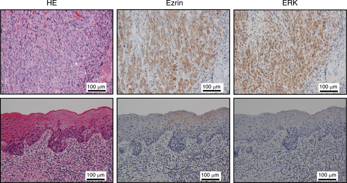
To screen for additional treatment targets against tongue cancer, we evaluated the contributions of extracellular signal-related kinase (ERK), AKT and ezrin in cancer development. Immunohistochemical staining showed that ERK and ezrin expressions were significantly higher in invasive squamous cell carcinoma than in carcinoma in situ. To investigate the roles of ERK and ezrin in cancer development, we used the non-woven silica fibre sheet CellbedTM with a structure resembling the loose connective tissue morphology in a novel 3D culture system. We confirmed that the 3D system using CellbedTM accurately mimicked cancer cell morphology in vivo. Furthermore, cell projections were much more apparent in 3D-cultured tongue cancer cell lines than in 2D cultures. Typically, under conventional 2D culture conditions, F-actin and cortactin are colocalized in the form of puncta within cells. However, in the 3D-cultured cells, colocalization was mainly observed at the cell margins, including the projections. Projections containing F-actin and cortactin colocalization were predicted to be invadopodia. Although suppressing ezrin expression with small interfering RNA transfection caused no marked changes in morphology, cell projection formation was decreased, and the tumour thickness in vertical sections after 3D culture was markedly decreased after suppressing ERK activity because both the invasion ability and proliferation were inhibited. An association between cortactin activation as well as ERK activity and invadopodia formation was detected. Our novel 3D culture systems using Cellbed™ are simple and useful for in vitro studies before conducting animal experiments. ERK contributes to tongue cancer development by increasing both cancer cell proliferation and migration via cortactin activation.
中文翻译:
在基于硅酸盐纤维的新型3D细胞培养系统中,ERK磷酸化作用在舌癌细胞的侵袭伪足形成中发挥作用
为了筛选针对舌癌的其他治疗靶标,我们评估了细胞外信号相关激酶(ERK),AKT和ezrin在癌症发展中的作用。免疫组织化学染色显示,浸润性鳞状细胞癌中ERK和ezrin的表达明显高于原位癌。为了研究ERK和ezrin在癌症发展中的作用,我们在新型3D培养系统中使用了具有类似于松散结缔组织形态的结构的无纺硅胶纤维片Cellbed TM。我们确认使用Cellbed TM的3D系统准确地模仿体内的癌细胞形态。此外,与2D培养相比,在3D培养的舌癌细胞系中细胞投射更为明显。通常,在常规的2D培养条件下,F-肌动蛋白和cortactin以点状的形式共定位在细胞内。但是,在3D培养的细胞中,共定位主要在细胞边缘(包括投影)处观察到。预测含有F-肌动蛋白和cortactin共定位的投影物是invadopodia。尽管用小的干扰RNA转染抑制ezrin表达不会引起形态上的显着变化,但是抑制ERK活性后,由于抑制了侵袭能力和增殖,细胞突起形成减少了,并且3D培养后垂直切片中的肿瘤厚度明显减少了。检测到了cortactin激活以及ERK活性与侵袭伪足形成之间的关联。我们使用Cellbed™的新型3D培养系统非常简单,可用于进行动物实验之前的体外研究。ERK通过增加癌细胞的增殖和通过cortactin激活的迁移来促进舌癌的发展。


 京公网安备 11010802027423号