Analytical and Bioanalytical Chemistry ( IF 3.8 ) Pub Date : 2019-01-25 , DOI: 10.1007/s00216-019-01577-9 Chunhuan Jiang , Ying Wang , Wei Song , Lehui Lu
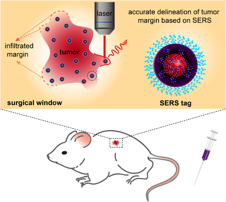
The failure of complete tumor resection during cancer surgery is a leading cause of lethal recurrence and metastasis. However, achieving accurate delineation of tumor margins intraoperatively remains extremely difficult because the infiltrated nature of a tumor usually gives an obscure margin and spreading microtumors. Recent studies show that surface-enhanced Raman scattering (SERS) has the potential to depict precisely the actual tumor extent with high sensitivity, specificity, and spatial resolution; thus providing a promising platform to improve the therapeutic efficiency. In this review, we discuss the recent progress in the use of SERS spectroscopy for intraoperative image-guided resection. We highlight key successes in the development of SERS tags and give insights into the design mechanism of rational SERS tags. We also discuss how to improve the performance of intraoperative navigation based on SERS and explore the challenges and future opportunities for the development of a more effective SERS-based platform.
ᅟ
中文翻译:
用术中表面增强拉曼光谱仪描绘肿瘤边缘
癌症手术期间无法完全切除肿瘤是致死性复发和转移的主要原因。然而,在术中实现肿瘤边界的精确描绘仍然非常困难,因为肿瘤的浸润性质通常会产生模糊的边界和散布的微瘤。最近的研究表明,表面增强拉曼散射(SERS)具有以高灵敏度,特异性和空间分辨率精确描绘实际肿瘤范围的潜力。从而为提高治疗效率提供了有希望的平台。在这篇综述中,我们讨论了在术中图像引导切除术中使用SERS光谱学的最新进展。我们重点介绍了SERS标签开发中的主要成功,并深入介绍了合理的SERS标签的设计机制。
ᅟ


 京公网安备 11010802027423号