Our official English website, www.x-mol.net, welcomes your
feedback! (Note: you will need to create a separate account there.)
Scanless volumetric imaging by selective access multifocal multiphoton microscopy.
Optica ( IF 8.4 ) Pub Date : 2019-01-20 , DOI: 10.1364/optica.6.000076 Yi Xue 1, 2 , Kalen P Berry 3 , Josiah R Boivin 4 , Christopher J Rowlands 5 , Yu Takiguchi 2, 6 , Elly Nedivi 3, 4, 7 , Peter T C So 1, 2, 5
Optica ( IF 8.4 ) Pub Date : 2019-01-20 , DOI: 10.1364/optica.6.000076 Yi Xue 1, 2 , Kalen P Berry 3 , Josiah R Boivin 4 , Christopher J Rowlands 5 , Yu Takiguchi 2, 6 , Elly Nedivi 3, 4, 7 , Peter T C So 1, 2, 5
Affiliation
|
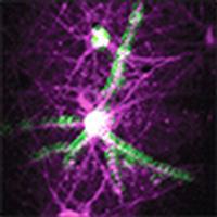
Simultaneous, high-resolution imaging across a large number of synaptic and dendritic sites is critical for understanding how neurons receive and integrate signals. Yet, functional imaging that targets a large number of submicrometer-sized synaptic and dendritic locations poses significant technical challenges. We demonstrate a new parallelized approach to address such questions, increasing the signal-to-noise ratio by an order of magnitude compared to previous approaches. This selective access multifocal multiphoton microscopy uses a spatial light modulator to generate multifocal excitation in three dimensions (3D) and a Gaussian-Laguerre phase plate to simultaneously detect fluorescence from these spots throughout the volume. We test the performance of this system by simultaneously recording Ca2+ dynamics from cultured neurons at 98-118 locations distributed throughout a 3D volume. This is the first demonstration of 3D imaging in a "single shot" and permits synchronized monitoring of signal propagation across multiple different dendrites.
中文翻译:

通过选择性访问多焦点多光子显微镜进行无扫描体积成像。
大量突触和树突位点的同步高分辨率成像对于理解神经元如何接收和整合信号至关重要。然而,针对大量亚微米大小的突触和树突位置的功能成像提出了重大的技术挑战。我们展示了一种新的并行方法来解决此类问题,与以前的方法相比,信噪比提高了一个数量级。这种选择性多焦点多光子显微镜使用空间光调制器在三维 (3D) 中产生多焦点激发,并使用高斯-拉盖尔相位板同时检测整个体积中这些点的荧光。我们通过同时记录分布在 3D 体积中 98-118 个位置的培养神经元的 Ca2+ 动态来测试该系统的性能。这是“单次”3D 成像的首次演示,并允许同步监测多个不同树突上的信号传播。
更新日期:2019-01-22
中文翻译:
通过选择性访问多焦点多光子显微镜进行无扫描体积成像。
大量突触和树突位点的同步高分辨率成像对于理解神经元如何接收和整合信号至关重要。然而,针对大量亚微米大小的突触和树突位置的功能成像提出了重大的技术挑战。我们展示了一种新的并行方法来解决此类问题,与以前的方法相比,信噪比提高了一个数量级。这种选择性多焦点多光子显微镜使用空间光调制器在三维 (3D) 中产生多焦点激发,并使用高斯-拉盖尔相位板同时检测整个体积中这些点的荧光。我们通过同时记录分布在 3D 体积中 98-118 个位置的培养神经元的 Ca2+ 动态来测试该系统的性能。这是“单次”3D 成像的首次演示,并允许同步监测多个不同树突上的信号传播。


 京公网安备 11010802027423号