Our official English website, www.x-mol.net, welcomes your
feedback! (Note: you will need to create a separate account there.)
SERS-Based Quantification of PSMA in Tissue Microarrays Allows Effective Stratification of Patients with Prostate Cancer
ACS Omega ( IF 3.7 ) Pub Date : 2018-12-06 00:00:00 , DOI: 10.1021/acsomega.8b01839 Manjari Bhamidipati 1 , Geuntaek Lee 2 , Isaac Kim 2 , Laura Fabris 3
ACS Omega ( IF 3.7 ) Pub Date : 2018-12-06 00:00:00 , DOI: 10.1021/acsomega.8b01839 Manjari Bhamidipati 1 , Geuntaek Lee 2 , Isaac Kim 2 , Laura Fabris 3
Affiliation
|
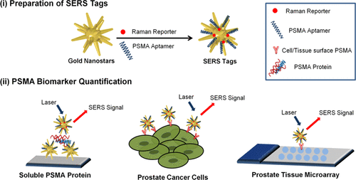
Prostate specific membrane antigen (PSMA), a type II membrane protein, is an attractive biomarker that has been validated clinically for the diagnosis of prostate cancer. In this study, we developed surface-enhanced Raman scattering (SERS) nanoprobes for PSMA detection and quantification at the single-cell level on prostate cancer cells. The cells were targeted employing SERS nanoprobes that consisted of gold nanostars functionalized with PSMA aptamer molecules. We were able to quantify picomolar concentrations of soluble PSMA protein and used the resulting calibration curve to estimate the expression of PSMA on the surface of the prostate cancer cell, LNCaP, at the single-cell level. Importantly, we employed these SERS tags to stratify prostate cancer patients by assessing PSMA expression in tissues contained in a prostate tissue microarray. The stratification results clearly correlated PSMA expression to recommended therapy groups, rendering the described method as an effective tool to aid in designing personalized therapeutic protocols. Benchmarking detection sensitivity against immunofluorescence staining and comparing stratification results obtained with the two methods allowed us to validate our novel approach against standard practices. On the basis of these results, we confirm the validity of PSMA as an effective biomarker for prostate cancer patient evaluation and propose SERS-based diagnostic techniques as integrative methods for the assessment of disease stage and the identification of effective therapeutic protocols.
中文翻译:

基于SERS的组织微阵列中PSMA的定量分析可对前列腺癌患者进行有效的分层
前列腺特异性膜抗原(PSMA)是一种II型膜蛋白,是一种有吸引力的生物标志物,已在临床上被证实可用于诊断前列腺癌。在这项研究中,我们开发了表面增强拉曼散射(SERS)纳米探针,用于在前列腺癌细胞的单细胞水平上进行PSMA检测和定量。使用SERS纳米探针靶向细胞,该探针由PSMA适体分子功能化的金纳米星组成。我们能够量化可溶性PSMA蛋白的皮摩尔浓度,并使用所得的校准曲线来估计PSMA在单细胞水平在前列腺癌细胞LNCaP上的表达。重要的是,我们通过评估前列腺组织微阵列中所含组织中的PSMA表达,将这些SERS标签用于对前列腺癌患者进行分层。分层结果清楚地将PSMA表达与推荐的治疗组相关联,从而使所描述的方法成为帮助设计个性化治疗方案的有效工具。对免疫荧光染色进行基准检测灵敏度比较,并比较两种方法获得的分层结果,这使我们能够对照标准方法验证我们的新方法。基于这些结果,我们确认了PSMA作为前列腺癌患者评估的有效生物标志物的有效性,并提出了基于SERS的诊断技术,作为评估疾病阶段和确定有效治疗方案的综合方法。将所描述的方法作为帮助设计个性化治疗方案的有效工具。对免疫荧光染色进行基准检测灵敏度比较,并比较两种方法获得的分层结果,这使我们能够对照标准方法验证我们的新方法。基于这些结果,我们确认了PSMA作为前列腺癌患者评估的有效生物标志物的有效性,并提出了基于SERS的诊断技术,作为评估疾病阶段和确定有效治疗方案的综合方法。将所描述的方法作为帮助设计个性化治疗方案的有效工具。对免疫荧光染色进行基准检测灵敏度比较,并比较两种方法获得的分层结果,这使我们能够对照标准方法验证我们的新方法。基于这些结果,我们确认了PSMA作为前列腺癌患者评估的有效生物标志物的有效性,并提出了基于SERS的诊断技术,作为评估疾病阶段和确定有效治疗方案的综合方法。
更新日期:2018-12-06
中文翻译:
基于SERS的组织微阵列中PSMA的定量分析可对前列腺癌患者进行有效的分层
前列腺特异性膜抗原(PSMA)是一种II型膜蛋白,是一种有吸引力的生物标志物,已在临床上被证实可用于诊断前列腺癌。在这项研究中,我们开发了表面增强拉曼散射(SERS)纳米探针,用于在前列腺癌细胞的单细胞水平上进行PSMA检测和定量。使用SERS纳米探针靶向细胞,该探针由PSMA适体分子功能化的金纳米星组成。我们能够量化可溶性PSMA蛋白的皮摩尔浓度,并使用所得的校准曲线来估计PSMA在单细胞水平在前列腺癌细胞LNCaP上的表达。重要的是,我们通过评估前列腺组织微阵列中所含组织中的PSMA表达,将这些SERS标签用于对前列腺癌患者进行分层。分层结果清楚地将PSMA表达与推荐的治疗组相关联,从而使所描述的方法成为帮助设计个性化治疗方案的有效工具。对免疫荧光染色进行基准检测灵敏度比较,并比较两种方法获得的分层结果,这使我们能够对照标准方法验证我们的新方法。基于这些结果,我们确认了PSMA作为前列腺癌患者评估的有效生物标志物的有效性,并提出了基于SERS的诊断技术,作为评估疾病阶段和确定有效治疗方案的综合方法。将所描述的方法作为帮助设计个性化治疗方案的有效工具。对免疫荧光染色进行基准检测灵敏度比较,并比较两种方法获得的分层结果,这使我们能够对照标准方法验证我们的新方法。基于这些结果,我们确认了PSMA作为前列腺癌患者评估的有效生物标志物的有效性,并提出了基于SERS的诊断技术,作为评估疾病阶段和确定有效治疗方案的综合方法。将所描述的方法作为帮助设计个性化治疗方案的有效工具。对免疫荧光染色进行基准检测灵敏度比较,并比较两种方法获得的分层结果,这使我们能够对照标准方法验证我们的新方法。基于这些结果,我们确认了PSMA作为前列腺癌患者评估的有效生物标志物的有效性,并提出了基于SERS的诊断技术,作为评估疾病阶段和确定有效治疗方案的综合方法。


 京公网安备 11010802027423号