Our official English website, www.x-mol.net, welcomes your
feedback! (Note: you will need to create a separate account there.)
Added Value of Microscale Raman Chemical Analysis in Mild Traumatic Brain Injury (TBI): A Comparison with Macroscale MRI
ACS Omega ( IF 3.7 ) Pub Date : 2018-12-06 00:00:00 , DOI: 10.1021/acsomega.8b02404 Dmitry Khalenkow , Sam Donche , Kim Braeckman , Christian Vanhove , Andre G. Skirtach
ACS Omega ( IF 3.7 ) Pub Date : 2018-12-06 00:00:00 , DOI: 10.1021/acsomega.8b02404 Dmitry Khalenkow , Sam Donche , Kim Braeckman , Christian Vanhove , Andre G. Skirtach

|
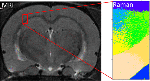
Diffuse axonal injury and microhemorrhages, common complications after mild traumatic brain injury (mTBI), can lead to neurodegeneration and disability and have negative socioeconomic consequences. Magnetic resonance imaging (MRI) is conventionally used to study brain injuries in vivo, but microscale damage common in mTBI is challenging to detect. Raman microscopy is an effective diagnostic tool to investigate cells and tissue in a label-free manner, but the scanning mode of Raman microscopy is typically used only in vitro. Here, we show that Raman microscopy complements in vivo MRI, providing the vital information on the structural and molecular changes caused by mTBI in rats. We demonstrate that a method based on Raman microscopy allows us to detect structural damage invisible by conventional MRI and spot molecular changes in protein/lipid concentrations caused by mild TBI.
中文翻译:

微型拉曼化学分析在轻度创伤性脑损伤(TBI)中的附加值:与大型MRI的比较
弥漫性轴索损伤和微出血是轻度颅脑损伤(mTBI)后的常见并发症,可导致神经退行性疾病和残疾,并对社会经济产生负面影响。传统上,磁共振成像(MRI)用于研究体内脑部损伤,但要检测出mTBI中常见的微尺度损伤是具有挑战性的。拉曼显微镜是一种无标记方式研究细胞和组织的有效诊断工具,但拉曼显微镜的扫描模式通常仅在体外使用。在这里,我们显示拉曼显微镜补充了体内MRI,提供了由mTBI在大鼠中引起的结构和分子变化的重要信息。
更新日期:2018-12-06
中文翻译:

微型拉曼化学分析在轻度创伤性脑损伤(TBI)中的附加值:与大型MRI的比较
弥漫性轴索损伤和微出血是轻度颅脑损伤(mTBI)后的常见并发症,可导致神经退行性疾病和残疾,并对社会经济产生负面影响。传统上,磁共振成像(MRI)用于研究体内脑部损伤,但要检测出mTBI中常见的微尺度损伤是具有挑战性的。拉曼显微镜是一种无标记方式研究细胞和组织的有效诊断工具,但拉曼显微镜的扫描模式通常仅在体外使用。在这里,我们显示拉曼显微镜补充了体内MRI,提供了由mTBI在大鼠中引起的结构和分子变化的重要信息。


 京公网安备 11010802027423号