当前位置:
X-MOL 学术
›
Neuropsychopharmacology
›
论文详情
Our official English website, www.x-mol.net, welcomes your
feedback! (Note: you will need to create a separate account there.)
Synaptic adaptations in the central amygdala and hypothalamic paraventricular nucleus associated with protracted ethanol abstinence in male rhesus monkeys.
Neuropsychopharmacology ( IF 6.6 ) Pub Date : 2018-12-05 , DOI: 10.1038/s41386-018-0290-7 V A Jimenez 1, 2 , M A Herman 3, 4 , V C Cuzon Carlson 1, 2 , N A Walter 2 , K A Grant 1, 2 , M Roberto 4
Neuropsychopharmacology ( IF 6.6 ) Pub Date : 2018-12-05 , DOI: 10.1038/s41386-018-0290-7 V A Jimenez 1, 2 , M A Herman 3, 4 , V C Cuzon Carlson 1, 2 , N A Walter 2 , K A Grant 1, 2 , M Roberto 4
Affiliation
|
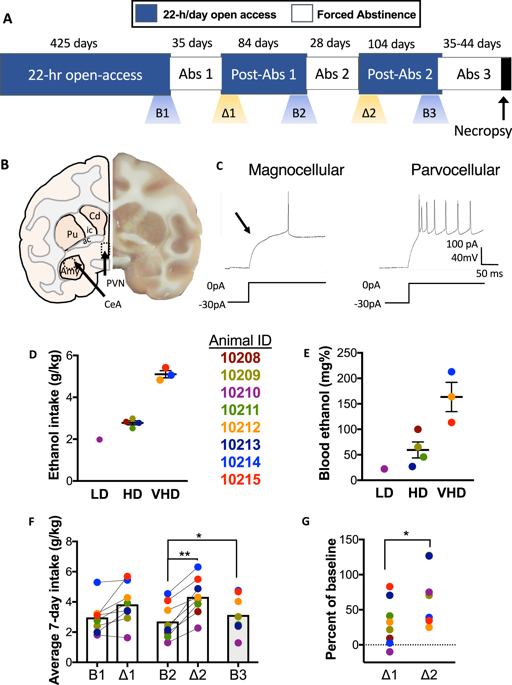
Alcohol use disorder is a significant global burden. Stress has been identified as an etiological factor in the initiation and continuation of ethanol consumption. Understanding adaptations within stress circuitry is an important step toward novel treatment strategies. The effects of protracted abstinence following long-term ethanol self-administration on the central nucleus of the amygdala (CeA) and the hypothalamic paraventricular nucleus (PVN) were evaluated in male rhesus monkeys. Using whole-cell patch-clamp electrophysiology, inhibitory GABAergic transmission in the CeA and excitatory glutamatergic transmission in the PVN were measured. CeA neurons from abstinent drinkers displayed an elevated baseline spontaneous inhibitory postsynaptic current (sIPSC) frequency compared with controls, indicating increased presynaptic GABA release. Application of acute ethanol significantly increased the frequency of sIPSCs in controls, but not in abstinent drinkers, suggesting a tolerance to ethanol-enhanced GABA release in abstinent rhesus monkeys with a history of chronic ethanol self-administration and repeated abstinence. In the PVN, the frequency of spontaneous excitatory postsynaptic currents (sEPSC) was elevated in abstinent drinkers compared with controls, indicating increased presynaptic glutamate release. Notably, acute ethanol decreased presynaptic glutamate release onto parvocellular PVN neurons in both controls and abstinent drinkers, suggesting a lack of tolerance to acute ethanol among PVN neurons. These results are the first to demonstrate distinct synaptic adaptations and ethanol sensitivity in both the extrahypothalamic and hypothalamic stress circuits in abstinent rhesus males. Importantly, our findings describe adaptations in stress circuitry present in the brain at a state during abstinence, just prior to relapse to ethanol drinking.
中文翻译:

中央杏仁核和下丘脑室旁核的突触适应与雄性恒河猴长期戒酒有关。
酒精使用障碍是一个重大的全球负担。压力已被确定为乙醇消耗开始和持续的病因学因素。了解压力回路中的适应是迈向新治疗策略的重要一步。在雄性恒河猴中评估长期戒酒后长期戒酒对杏仁核中央核 (CeA) 和下丘脑室旁核 (PVN) 的影响。使用全细胞膜片钳电生理学,测量了 CeA 中的抑制性 GABAergic 传输和 PVN 中的兴奋性谷氨酸能传输。与对照组相比,戒酒者的 CeA 神经元基线自发抑制性突触后电流 (sIPSC) 频率升高,表明突触前 GABA 释放增加。急性乙醇的应用显着增加了 sIPSCs 在对照组中的频率,但在戒酒者中没有,这表明在具有慢性乙醇自我给药和反复戒酒史的戒酒恒河猴中对乙醇增强的 GABA 释放具有耐受性。在 PVN 中,与对照组相比,戒酒者的自发兴奋性突触后电流 (sEPSC) 频率升高,表明突触前谷氨酸释放增加。值得注意的是,急性乙醇减少了突触前谷氨酸释放到对照组和戒酒者的细小细胞 PVN 神经元上,表明 PVN 神经元对急性乙醇缺乏耐受性。这些结果首次证明了戒断恒河猴雄性下丘脑外和下丘脑应激回路中明显的突触适应和乙醇敏感性。重要的是,我们的研究结果描述了在戒酒期间大脑中存在的压力回路的适应性,就在酒精饮用复发之前。
更新日期:2019-01-26
中文翻译:
中央杏仁核和下丘脑室旁核的突触适应与雄性恒河猴长期戒酒有关。
酒精使用障碍是一个重大的全球负担。压力已被确定为乙醇消耗开始和持续的病因学因素。了解压力回路中的适应是迈向新治疗策略的重要一步。在雄性恒河猴中评估长期戒酒后长期戒酒对杏仁核中央核 (CeA) 和下丘脑室旁核 (PVN) 的影响。使用全细胞膜片钳电生理学,测量了 CeA 中的抑制性 GABAergic 传输和 PVN 中的兴奋性谷氨酸能传输。与对照组相比,戒酒者的 CeA 神经元基线自发抑制性突触后电流 (sIPSC) 频率升高,表明突触前 GABA 释放增加。急性乙醇的应用显着增加了 sIPSCs 在对照组中的频率,但在戒酒者中没有,这表明在具有慢性乙醇自我给药和反复戒酒史的戒酒恒河猴中对乙醇增强的 GABA 释放具有耐受性。在 PVN 中,与对照组相比,戒酒者的自发兴奋性突触后电流 (sEPSC) 频率升高,表明突触前谷氨酸释放增加。值得注意的是,急性乙醇减少了突触前谷氨酸释放到对照组和戒酒者的细小细胞 PVN 神经元上,表明 PVN 神经元对急性乙醇缺乏耐受性。这些结果首次证明了戒断恒河猴雄性下丘脑外和下丘脑应激回路中明显的突触适应和乙醇敏感性。重要的是,我们的研究结果描述了在戒酒期间大脑中存在的压力回路的适应性,就在酒精饮用复发之前。


 京公网安备 11010802027423号