当前位置:
X-MOL 学术
›
J. Am. Soc. Echocardiog.
›
论文详情
Our official English website, www.x-mol.net, welcomes your
feedback! (Note: you will need to create a separate account there.)
Validation of a Holographic Display for Quantification of Mitral Annular Dynamics by Three-Dimensional Echocardiography.
Journal of the American Society of Echocardiography ( IF 5.4 ) Pub Date : 2018-10-05 , DOI: 10.1016/j.echo.2018.08.010 Karl-Andreas Dumont 1 , John-Peder Escobar Kvitting 1 , Jørn S Karlsen 2 , Espen W Remme 3 , John Hausken 4 , Runar Lundblad 1 , Arnt E Fiane 5 , Stig Urheim 6
Journal of the American Society of Echocardiography ( IF 5.4 ) Pub Date : 2018-10-05 , DOI: 10.1016/j.echo.2018.08.010 Karl-Andreas Dumont 1 , John-Peder Escobar Kvitting 1 , Jørn S Karlsen 2 , Espen W Remme 3 , John Hausken 4 , Runar Lundblad 1 , Arnt E Fiane 5 , Stig Urheim 6
Affiliation
|
BACKGROUND
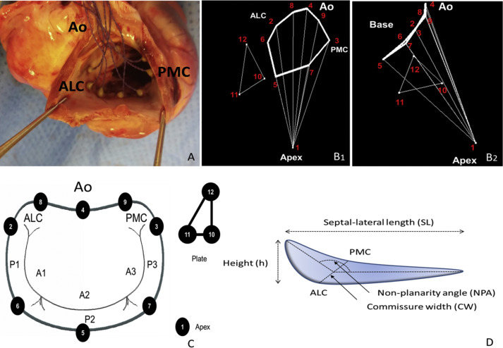
Three-dimensional (3D) echocardiography with multiplanar reconstruction (MPR) is used clinically to quantify the mitral annulus. MPR images are, however, presented on a two-dimensional screen, calling into question their accuracy. An alternative to MPR is an autostereoscopic holographic display that enables in-depth visualization of 3D echocardiographic data without the need for special glasses. The aim of this study was to validate an autostereoscopic display using sonomicrometry as a gold standard.
METHODS
In 11 anesthetized open-chest pigs, sonomicrometric crystals were placed along the mitral annulus and near the left ventricular apex. High-fidelity catheters measured left atrial and ventricular pressures. Adjustments of pre- and afterload were done by constriction of the inferior vena cava and the ascending aorta, respectively. Three-dimensional epicardial echocardiography was obtained from an apical view and converted to the autostereoscopic display. A 3D virtual semitransparent annular surface (VSAS) was generated to measure commissure width (CW), septal-lateral length, area of the mitral annular surface, nonplanarity angle, and the annular height-to-commissure width ratio in mid-systole and late diastole.
RESULTS
Mitral annular measurements from the 3D VSAS derived from the 3D echocardiographic images and autostereoscopic display correlated well with sonomicrometry over a range of loading conditions: CW length (r = 0.98, P < .00001), septal-lateral length (r = 0.98, P < .00001), annular surface area (r = 0.93, P < .001), nonplanarity angle (r = 0.87, P < .001), and annular height-to-commissure width ratio (r = 0.85, P < .01). The 3D VSAS showed better agreement with the sonomicrometric measurements compared with MPR.
CONCLUSIONS
Mitral annular measurements using 3D VSAS correlate well with sonomicrometry over a range of loading conditions and may represent a powerful tool for noninvasive quantification of mitral annular dynamics.
中文翻译:

通过三维超声心动图量化二尖瓣环动力学的全息显示的验证。
背景技术具有多平面重建(MPR)的三维(3D)超声心动图在临床上用于量化二尖瓣环。但是,MPR图像显示在二维屏幕上,这质疑了它们的准确性。MPR的替代产品是自动立体全息显示器,无需特殊眼镜即可实现3D超声心动图数据的深入可视化。这项研究的目的是验证使用体测法作为黄金标准的自动立体显示器。方法在11只麻醉的开胸猪中,沿二尖瓣环和左心室顶点附近放置体温晶体。高保真导管测量左心房和心室压力。分别通过下腔静脉和升主动脉的收缩来调节前后负荷。从心尖获得三维心外膜超声心动图,并将其转换为自动立体显示。生成了一个3D虚拟半透明环形表面(VSAS),以测量收缩中期和收缩后期的合缝宽度(CW),隔侧长,二尖瓣环形表面的面积,非平面角以及环形高度与合缝的宽度比。舒张期。结果从3D超声心动图图像和自动立体显示获得的3D VSAS的二尖瓣环测量值与在一系列载荷条件下的体测法具有很好的相关性:CW长度(r = 0.98,P <.00001),隔侧长度(r = 0.98, P <.00001),环形表面积(r = 0.93,P <.001),非平面角(r = 0.87,P <.001)和环形高度与合缝宽度之比(r = 0.85,P <。 01)。与MPR相比,3D VSAS与体测法测量显示出更好的一致性。结论使用3D VSAS进行的二尖瓣环测量在一定的负荷条件下与体模测量法具有很好的相关性,并且可能代表了用于二尖瓣环动力学的非侵入性量化的强大工具。
更新日期:2018-10-05
中文翻译:
通过三维超声心动图量化二尖瓣环动力学的全息显示的验证。
背景技术具有多平面重建(MPR)的三维(3D)超声心动图在临床上用于量化二尖瓣环。但是,MPR图像显示在二维屏幕上,这质疑了它们的准确性。MPR的替代产品是自动立体全息显示器,无需特殊眼镜即可实现3D超声心动图数据的深入可视化。这项研究的目的是验证使用体测法作为黄金标准的自动立体显示器。方法在11只麻醉的开胸猪中,沿二尖瓣环和左心室顶点附近放置体温晶体。高保真导管测量左心房和心室压力。分别通过下腔静脉和升主动脉的收缩来调节前后负荷。从心尖获得三维心外膜超声心动图,并将其转换为自动立体显示。生成了一个3D虚拟半透明环形表面(VSAS),以测量收缩中期和收缩后期的合缝宽度(CW),隔侧长,二尖瓣环形表面的面积,非平面角以及环形高度与合缝的宽度比。舒张期。结果从3D超声心动图图像和自动立体显示获得的3D VSAS的二尖瓣环测量值与在一系列载荷条件下的体测法具有很好的相关性:CW长度(r = 0.98,P <.00001),隔侧长度(r = 0.98, P <.00001),环形表面积(r = 0.93,P <.001),非平面角(r = 0.87,P <.001)和环形高度与合缝宽度之比(r = 0.85,P <。 01)。与MPR相比,3D VSAS与体测法测量显示出更好的一致性。结论使用3D VSAS进行的二尖瓣环测量在一定的负荷条件下与体模测量法具有很好的相关性,并且可能代表了用于二尖瓣环动力学的非侵入性量化的强大工具。


 京公网安备 11010802027423号