当前位置:
X-MOL 学术
›
npj Schizophr.
›
论文详情
Our official English website, www.x-mol.net, welcomes your
feedback! (Note: you will need to create a separate account there.)
Medial septum activation produces opposite effects on dopamine neuron activity in the ventral tegmental area and substantia nigra in MAM vs. normal rats.
npj Schizophrenia ( IF 5.7 ) Pub Date : 2018-09-03 , DOI: 10.1038/s41537-018-0059-3 David M Bortz 1, 2, 3 , Anthony A Grace 1, 2, 3
npj Schizophrenia ( IF 5.7 ) Pub Date : 2018-09-03 , DOI: 10.1038/s41537-018-0059-3 David M Bortz 1, 2, 3 , Anthony A Grace 1, 2, 3
Affiliation
|
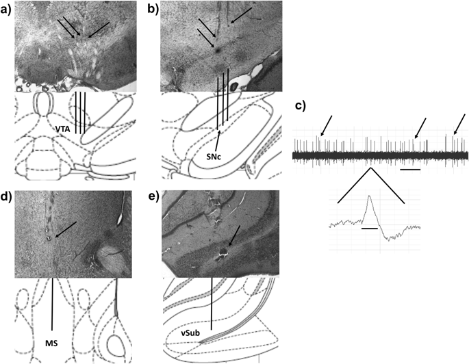
The medial septum (MS) differentially impacts midbrain dopamine (DA) neuron activity via the ventral hippocampus, a region implicated in DA-related disorders. However, whether MS regulation of ventral tegmental area (VTA) and substantia nigra pars compacta (SNc) is disrupted in a developmental disruption model of schizophrenia is unknown. Male Sprague-Dawley rats were exposed at gestational day 17 to methylazoxymethanol (MAM) or saline. As adults, NMDA (0.75 µg/0.2 µL) was infused into the MS, and either DA neuron activity in the VTA and SNc (7-9 anesthetized rats per group) or amphetamine-induced hyperlocomotion (AIH, 11-13 rats per group) was measured. MS activation produced a 58% increase in the number of spontaneously active DA neurons in VTA and a 37% decrease in SNc in saline rats. However, MS activation produced opposite effects on DA population activity in MAM rats, decreasing VTA DA activity by 51% and increasing SNc DA activity by 47%. MS activation also increased AIH by 113% in MAM rats, opposite of what is seen in intact rats. The effect in behavioral output may be due to disrupted GABAergic regulation of SNc as bicuculline infusion into vSub, which selectively prevented the MS activation-induced decrease in SNc DA activity in intact rats, prevented the increase in AIH and SNc DA activity in MAM rats. These findings demonstrate that the regulation of midbrain DA neurons by the MS is disrupted in this well-validated animal model, suggesting that it could be a potential locus for pharmacological intervention in disorders such as schizophrenia.
中文翻译:

与正常大鼠相比,MAM 的内侧隔膜激活对腹侧被盖区和黑质的多巴胺神经元活性产生相反的影响。
内侧隔膜 (MS) 通过腹侧海马(与 DA 相关疾病有关的区域)对中脑多巴胺 (DA) 神经元活动产生差异性影响。然而,在精神分裂症的发育破坏模型中,腹侧被盖区 (VTA) 和黑质致密部 (SNc) 的 MS 调节是否受到破坏尚不清楚。雄性 Sprague-Dawley 大鼠在妊娠第 17 天暴露于甲基偶氮甲醇 (MAM) 或盐水。成年后,将 NMDA (0.75 µg/0.2 µL) 注入 MS,并观察 VTA 和 SNc 中的 DA 神经元活性(每组 7-9 只麻醉大鼠)或安非他明诱导的过度运动(AIH,每组 11-13 只大鼠) ) 进行了测量。在盐水大鼠中,MS 激活使 VTA 中自发活跃的 DA 神经元数量增加 58%,SNc 减少 37%。然而,MS 激活对 MAM 大鼠的 DA 群体活性产生相反的影响,使 VTA DA 活性降低 51%,使 SNc DA 活性增加 47%。 MS 激活还使 MAM 大鼠的 AIH 增加了 113%,这与在完整大鼠中观察到的相反。行为输出的影响可能是由于荷包牡丹碱输注到 vSub 中时,SNc 的 GABA 能调节被破坏,选择性地阻止了 MS 激活诱导的完整大鼠 SNc DA 活性的降低,阻止了 MAM 大鼠中 AIH 和 SNc DA 活性的增加。这些发现表明,在这种经过充分验证的动物模型中,MS 对中脑 DA 神经元的调节受到破坏,这表明它可能是对精神分裂症等疾病进行药物干预的潜在场所。
更新日期:2018-09-03
中文翻译:
与正常大鼠相比,MAM 的内侧隔膜激活对腹侧被盖区和黑质的多巴胺神经元活性产生相反的影响。
内侧隔膜 (MS) 通过腹侧海马(与 DA 相关疾病有关的区域)对中脑多巴胺 (DA) 神经元活动产生差异性影响。然而,在精神分裂症的发育破坏模型中,腹侧被盖区 (VTA) 和黑质致密部 (SNc) 的 MS 调节是否受到破坏尚不清楚。雄性 Sprague-Dawley 大鼠在妊娠第 17 天暴露于甲基偶氮甲醇 (MAM) 或盐水。成年后,将 NMDA (0.75 µg/0.2 µL) 注入 MS,并观察 VTA 和 SNc 中的 DA 神经元活性(每组 7-9 只麻醉大鼠)或安非他明诱导的过度运动(AIH,每组 11-13 只大鼠) ) 进行了测量。在盐水大鼠中,MS 激活使 VTA 中自发活跃的 DA 神经元数量增加 58%,SNc 减少 37%。然而,MS 激活对 MAM 大鼠的 DA 群体活性产生相反的影响,使 VTA DA 活性降低 51%,使 SNc DA 活性增加 47%。 MS 激活还使 MAM 大鼠的 AIH 增加了 113%,这与在完整大鼠中观察到的相反。行为输出的影响可能是由于荷包牡丹碱输注到 vSub 中时,SNc 的 GABA 能调节被破坏,选择性地阻止了 MS 激活诱导的完整大鼠 SNc DA 活性的降低,阻止了 MAM 大鼠中 AIH 和 SNc DA 活性的增加。这些发现表明,在这种经过充分验证的动物模型中,MS 对中脑 DA 神经元的调节受到破坏,这表明它可能是对精神分裂症等疾病进行药物干预的潜在场所。


 京公网安备 11010802027423号