Journal of the American Society of Echocardiography ( IF 6.5 ) Pub Date : 2018-08-23 , DOI: 10.1016/j.echo.2018.07.010 Yu Wang , Miao Fan , Faiza Amber Siddiqui , Meilian Wang , Wei Sun , Xue Sun , Wenjia Lei , Ying Zhang
|
Background
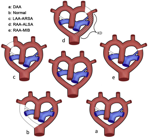
Obtaining an accurate diagnosis of fetal aortic arch anomalies is often difficult with traditional two-dimensional (2D) sonography. Thus, the aim of this study was to assess the value of three-dimensional (3D) sonography with spatiotemporal image correlation for diagnosing fetal aortic arch anomalies using a novel algorithm for 3D volume analysis.
Methods
Two-dimensional and 3D echocardiographic image data from 300 normal fetuses and 30 fetuses with aortic arch anomalies were retrospectively reviewed. Grayscale and high-definition flow imaging data were available for 2D echocardiography. Three-dimensional volumes were acquired in parasagittal views with high-definition flow imaging information. Volume postprocessing was performed using a novel algorithm to obtain 3D tomographic ultrasound imaging slices and color-rendered images. Detection of aortic arch positions, aberrant brachiocephalic patterns, and aortic arch anomalies was compared for 2D and 3D modalities. Postnatal echocardiography was used as the truth standard in assessing aortic anatomy.
Results
In total, four cases of double aortic arch, 21 cases of right aortic arch, one case of retroesophageal aortic arch, and four cases of left aortic arch with aberrant right subclavian arteries were included. Inter- and intraobserver agreement were excellent for 2D and 3D modalities. The 3D modality offered better sensitivity and accuracy compared with 2D imaging for the detection of brachiocephalic anomalies (P < .01) and arch anomalies (P < .01) but comparable sensitivity for arch position. There was no difference in specificity for both modalities.
Conclusions
The proposed novel algorithm for volume postprocessing ensures that 3D reconstructed images are obtained with high repeatability. The addition of 3D spatiotemporal image correlation to fetal echocardiography may offer a more accurate diagnosis for arch anomalies.
中文翻译:
胎儿主动脉弓畸形的准确诊断策略:时空图像相关的三维超声检查和体积分析新算法的好处
背景
传统的二维(2D)超声检查通常很难获得胎儿主动脉弓畸形的准确诊断。因此,本研究的目的是使用一种新颖的3D体积分析算法,评估具有时空图像相关性的三维(3D)超声检查对诊断胎儿主动脉弓畸形的价值。
方法
回顾性分析了300例正常胎儿和30例主动脉弓畸形胎儿的二维和3D超声心动图图像数据。灰度和高清流成像数据可用于2D超声心动图。在矢状旁观中获取具有三维流成像信息的三维体。使用新型算法执行体积后处理,以获得3D断层扫描超声成像切片和彩色渲染图像。比较了2D和3D模式的主动脉弓位置,异常头臂畸形和主动脉弓异常的检测。产后超声心动图被用作评估主动脉解剖结构的真实标准。
结果
总共包括双主动脉弓4例,右主动脉弓21例,食管后主动脉弓1例,右锁骨下动脉异常的左主动脉弓4例。观察者之间和观察者内部的协议非常适合2D和3D模式。与2D成像相比,3D模态检测肱头畸形(P <.01)和足弓异常(P <.01)的灵敏度和准确性更高,但对足弓位置的灵敏度却相当。两种方式的特异性没有差异。
结论
所提出的用于体积后处理的新颖算法确保获得具有高可重复性的3D重建图像。将3D时空图像相关性添加到胎儿超声心动图可以为弓畸形提供更准确的诊断。


 京公网安备 11010802027423号