当前位置:
X-MOL 学术
›
Neurobiol. Learn. Mem.
›
论文详情
Our official English website, www.x-mol.net, welcomes your
feedback! (Note: you will need to create a separate account there.)
Cerebellar injury and impaired function in a rabbit model of maternal inflammation induced neonatal brain injury.
Neurobiology of Learning and Memory ( IF 2.2 ) Pub Date : 2018-07-24 , DOI: 10.1016/j.nlm.2018.07.005 Zhi Zhang 1 , Shilpa Narayan 1 , Lilly Su 1 , Hanaa Al-Alawyat 1 , Jinhuan Liu 1 , Sujatha Kannan 1
Neurobiology of Learning and Memory ( IF 2.2 ) Pub Date : 2018-07-24 , DOI: 10.1016/j.nlm.2018.07.005 Zhi Zhang 1 , Shilpa Narayan 1 , Lilly Su 1 , Hanaa Al-Alawyat 1 , Jinhuan Liu 1 , Sujatha Kannan 1
Affiliation
|
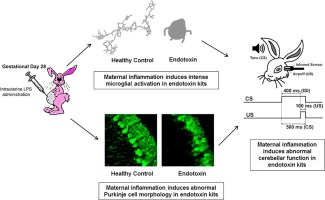
Cerebellum is involved in higher cognitive functions and plays important roles in neurological disorders. Cerebellar injury has been detected frequently in patients with preterm birth resulting in cognitive dysfunction later in life. Maternal infection and inflammation is associated with preterm birth and in neonatal brain injury. We have previously shown that intrauterine lipopolysaccharide (LPS) exposure induces white matter injury and microglial activation in the cerebral white matter tracts of neonatal rabbits, resulting in motor deficits consistent with the clinical findings of cerebral palsy (CP). Here we investigated whether intrauterine LPS exposure induced cerebellar inflammation and functional impairment. Timed-pregnant New Zealand white rabbits underwent a laparotomy on gestational day 28 (G28) and LPS (3200 EU, endotoxin group) was injected along the wall of the uterus as previously described. Controls did not receive surgical intervention. Kits born to control and endotoxin treated dams were euthanized on postnatal day (PND)1 (3 days post-injury) or PND5 (7 days post-injury) and cerebellum evaluated for presence of inflammation. The microglial morphology in cerebellar white matter areas was analyzed using Neurolucida and Neurolucida Explorer. mRNA expression of inflammatory cytokines was quantified by real-time-PCR. We found that intrauterine exposure to LPS induced intensive microglial activation in cerebellar white matter areas, as evidenced by increased numbers of activated microglia and morphological changes (amoeboid soma and retracted processes) that was accompanied by significant increases in pro-inflammatory cytokines. The Purkinje cell layer was less developed in endotoxin exposed kits than healthy controls. In kits that survived to PND 60, soma size and cell density of Purkinje cells were significantly decreased in endotoxin exposed kits compared to controls. The findings of altered Purkinje cell morphology were consistent with impaired cerebellar function as tested by eye-blink conditioning at 1 month of age. The results indicate that the cerebellum is vulnerable to perinatal insults and that therapies targeting cerebellar inflammation and injury may help in improving outcomes and function.
中文翻译:

在孕产妇炎症引起的新生儿脑损伤的兔子模型中,小脑损伤和功能受损。
小脑参与较高的认知功能,并在神经系统疾病中起重要作用。早产患者经常检测到小脑损伤,导致以后的生活中出现认知功能障碍。产妇感染和炎症与早产和新生儿脑损伤有关。先前我们已经证明,宫内脂多糖(LPS)暴露会引起新生兔脑白质束中的白质损伤和小胶质细胞活化,从而导致运动障碍,与脑瘫(CP)的临床表现相符。在这里,我们调查了宫内LPS暴露是否引起小脑发炎和功能障碍。定时妊娠的新西兰白兔在妊娠第28天(G28)和LPS(3200 EU,如前所述,沿子宫壁注射内毒素组)。对照组未接受手术干预。在出生后第1天(PND)1(受伤后3天)或PND5(受伤后7天)对对照组和经内毒素处理的大坝出生的试剂盒实施安乐死,并评估小脑是否存在炎症。使用Neurolucida和Neurolucida Explorer分析小脑白质区域的小胶质细胞形态。通过实时PCR定量炎性细胞因子的mRNA表达。我们发现,子宫内暴露于LPS会在小脑白质区域引起强烈的小胶质细胞活化,这由活化的小胶质细胞数量增加和形态变化(变形肌体和缩回过程)所伴生,同时伴随着促炎性细胞因子的显着增加。暴露于内毒素的试剂盒中的浦肯野细胞层比健康对照少。与对照组相比,在暴露至PND 60的试剂盒中,暴露于内毒素的试剂盒中浦肯野细胞的体大小和细胞密度显着降低。浦肯野细胞形态改变的发现与小脑功能受损在1个月大时通过眨眼条件进行了测试。结果表明,小脑易受围产期伤害,针对小脑炎症和损伤的疗法可能有助于改善预后和功能。浦肯野细胞形态改变的发现与小脑功能受损在1个月大时通过眨眼条件进行了测试。结果表明,小脑易受围产期伤害,针对小脑炎症和损伤的疗法可能有助于改善预后和功能。浦肯野细胞形态改变的发现与小脑功能受损在1个月大时通过眨眼条件进行了测试。结果表明,小脑易受围产期伤害,针对小脑炎症和损伤的疗法可能有助于改善预后和功能。
更新日期:2018-07-24
中文翻译:
在孕产妇炎症引起的新生儿脑损伤的兔子模型中,小脑损伤和功能受损。
小脑参与较高的认知功能,并在神经系统疾病中起重要作用。早产患者经常检测到小脑损伤,导致以后的生活中出现认知功能障碍。产妇感染和炎症与早产和新生儿脑损伤有关。先前我们已经证明,宫内脂多糖(LPS)暴露会引起新生兔脑白质束中的白质损伤和小胶质细胞活化,从而导致运动障碍,与脑瘫(CP)的临床表现相符。在这里,我们调查了宫内LPS暴露是否引起小脑发炎和功能障碍。定时妊娠的新西兰白兔在妊娠第28天(G28)和LPS(3200 EU,如前所述,沿子宫壁注射内毒素组)。对照组未接受手术干预。在出生后第1天(PND)1(受伤后3天)或PND5(受伤后7天)对对照组和经内毒素处理的大坝出生的试剂盒实施安乐死,并评估小脑是否存在炎症。使用Neurolucida和Neurolucida Explorer分析小脑白质区域的小胶质细胞形态。通过实时PCR定量炎性细胞因子的mRNA表达。我们发现,子宫内暴露于LPS会在小脑白质区域引起强烈的小胶质细胞活化,这由活化的小胶质细胞数量增加和形态变化(变形肌体和缩回过程)所伴生,同时伴随着促炎性细胞因子的显着增加。暴露于内毒素的试剂盒中的浦肯野细胞层比健康对照少。与对照组相比,在暴露至PND 60的试剂盒中,暴露于内毒素的试剂盒中浦肯野细胞的体大小和细胞密度显着降低。浦肯野细胞形态改变的发现与小脑功能受损在1个月大时通过眨眼条件进行了测试。结果表明,小脑易受围产期伤害,针对小脑炎症和损伤的疗法可能有助于改善预后和功能。浦肯野细胞形态改变的发现与小脑功能受损在1个月大时通过眨眼条件进行了测试。结果表明,小脑易受围产期伤害,针对小脑炎症和损伤的疗法可能有助于改善预后和功能。浦肯野细胞形态改变的发现与小脑功能受损在1个月大时通过眨眼条件进行了测试。结果表明,小脑易受围产期伤害,针对小脑炎症和损伤的疗法可能有助于改善预后和功能。


 京公网安备 11010802027423号