当前位置:
X-MOL 学术
›
J. Control. Release
›
论文详情
Our official English website, www.x-mol.net, welcomes your
feedback! (Note: you will need to create a separate account there.)
Photodynamic cancer therapy enhances accumulation of nanoparticles in tumor-associated myeloid cells.
Journal of Controlled Release ( IF 10.5 ) Pub Date : 2019-12-31 , DOI: 10.1016/j.jconrel.2019.12.052 Ruben V Huis In 't Veld 1 , Laila Ritsma 2 , Jan Willem Kleinovink 3 , Ivo Que 1 , Ferry Ossendorp 4 , Luis J Cruz 1
Journal of Controlled Release ( IF 10.5 ) Pub Date : 2019-12-31 , DOI: 10.1016/j.jconrel.2019.12.052 Ruben V Huis In 't Veld 1 , Laila Ritsma 2 , Jan Willem Kleinovink 3 , Ivo Que 1 , Ferry Ossendorp 4 , Luis J Cruz 1
Affiliation
|
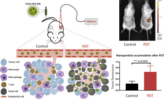
In cancer treatment, nanomedicines may be employed in an attempt to improve the tumor localization of antineoplastic drugs e.g. immunotherapeutic agents either through passive or active targeting, thereby potentially enhancing therapeutic effect and reducing undesired off-target effects. However, a large number of administrated nanocarriers often fail to reach the tumor area. In the present study, we show that photodynamic therapy (PDT) enhances the tumor accumulation of systemically administered lipid-PEG layer coated poly (lactic-co-glycolic acid) (PLGA) nanoparticles (NP). Intravital microscopy and histological analysis of the tumor area reveal that the tumor vasculature was disrupted after PDT, disturbing blood flow and coinciding with entrapment of nanocarriers in the tumor area. We observed that the nanoparticles accumulating after treatment do not confine to specific locations within the tumor, but rather localize to various cells present throughout the tumor area. Finally, we show by flow cytometry that NP accumulation occurred mostly in immune cells of the myeloid lineage present in the tumor microenvironment (TME) as well as in tumor cells, albeit to a lower extent. These data expose opportunities for combination treatments of clinical PDT with NP-based immunotherapy to modulate the TME and improve antitumor immune responses.
中文翻译:

光动力癌症疗法可增强纳米颗粒在肿瘤相关髓样细胞中的积累。
在癌症治疗中,可以采用纳米药物来尝试通过被动或主动靶向来改善抗肿瘤药物(例如免疫治疗剂)在肿瘤中的定位,从而潜在地增强治疗效果并降低不良的脱靶作用。然而,大量施用的纳米载体常常不能到达肿瘤区域。在本研究中,我们表明光动力疗法(PDT)增强了全身给药脂质-PEG涂层包裹的聚乳酸-乙醇酸(PLGA)纳米颗粒(NP)的肿瘤蓄积。活体显微镜检查和对肿瘤区域的组织学分析表明,PDT后肿瘤血管系统被破坏,扰乱了血流并与纳米载体在肿瘤区域中的滞留相吻合。我们观察到,治疗后积累的纳米颗粒并不局限于肿瘤内的特定位置,而是位于整个肿瘤区域内的各种细胞内。最后,我们通过流式细胞仪显示,NP积累主要发生在肿瘤微环境(TME)中存在的髓系谱系的免疫细胞以及肿瘤细胞中,尽管程度较低。这些数据揭示了临床PDT与基于NP的免疫疗法联合治疗以调节TME并改善抗肿瘤免疫反应的机会。我们通过流式细胞仪显示,NP积累主要发生在肿瘤微环境(TME)中存在的髓系谱系的免疫细胞以及肿瘤细胞中,尽管程度较低。这些数据揭示了临床PDT与基于NP的免疫疗法联合治疗以调节TME并改善抗肿瘤免疫反应的机会。我们通过流式细胞仪显示,NP积累主要发生在肿瘤微环境(TME)中存在的髓系谱系的免疫细胞以及肿瘤细胞中,尽管程度较低。这些数据揭示了临床PDT与基于NP的免疫疗法联合治疗以调节TME并改善抗肿瘤免疫反应的机会。
更新日期:2019-12-31
中文翻译:
光动力癌症疗法可增强纳米颗粒在肿瘤相关髓样细胞中的积累。
在癌症治疗中,可以采用纳米药物来尝试通过被动或主动靶向来改善抗肿瘤药物(例如免疫治疗剂)在肿瘤中的定位,从而潜在地增强治疗效果并降低不良的脱靶作用。然而,大量施用的纳米载体常常不能到达肿瘤区域。在本研究中,我们表明光动力疗法(PDT)增强了全身给药脂质-PEG涂层包裹的聚乳酸-乙醇酸(PLGA)纳米颗粒(NP)的肿瘤蓄积。活体显微镜检查和对肿瘤区域的组织学分析表明,PDT后肿瘤血管系统被破坏,扰乱了血流并与纳米载体在肿瘤区域中的滞留相吻合。我们观察到,治疗后积累的纳米颗粒并不局限于肿瘤内的特定位置,而是位于整个肿瘤区域内的各种细胞内。最后,我们通过流式细胞仪显示,NP积累主要发生在肿瘤微环境(TME)中存在的髓系谱系的免疫细胞以及肿瘤细胞中,尽管程度较低。这些数据揭示了临床PDT与基于NP的免疫疗法联合治疗以调节TME并改善抗肿瘤免疫反应的机会。我们通过流式细胞仪显示,NP积累主要发生在肿瘤微环境(TME)中存在的髓系谱系的免疫细胞以及肿瘤细胞中,尽管程度较低。这些数据揭示了临床PDT与基于NP的免疫疗法联合治疗以调节TME并改善抗肿瘤免疫反应的机会。我们通过流式细胞仪显示,NP积累主要发生在肿瘤微环境(TME)中存在的髓系谱系的免疫细胞以及肿瘤细胞中,尽管程度较低。这些数据揭示了临床PDT与基于NP的免疫疗法联合治疗以调节TME并改善抗肿瘤免疫反应的机会。


 京公网安备 11010802027423号