Our official English website, www.x-mol.net, welcomes your
feedback! (Note: you will need to create a separate account there.)
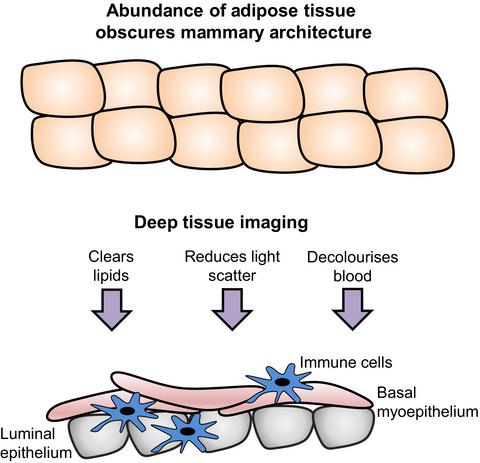
Deep imaging reveals new insights into mammary gland architecture and breast cancer susceptibility.
The FEBS Journal ( IF 5.5 ) Pub Date : 2019-12-26 , DOI: 10.1111/febs.15165 Wendy V Ingman 1, 2
The FEBS Journal ( IF 5.5 ) Pub Date : 2019-12-26 , DOI: 10.1111/febs.15165 Wendy V Ingman 1, 2
Affiliation
|
The abundance of adipose tissue in the mammary gland obscures vision of the 3-dimensional architecture. Hitchcock et al. employed a new technique of deep tissue imaging that has enabled visualisation of dynamic interactions between mammary gland epithelial and immune cells with unprecedented 3-dimensional clarity. Deep imaging will help further our understanding of the complex biological interactions that underpin both normal mammary gland development and the susceptibility of this tissue to cancer. This knowledge will assist the development of much-needed prevention strategies to reduce the incidence of breast cancer. Comment on: https://doi.org/10.1111/febs.15126.
中文翻译:

深度成像揭示了对乳腺结构和乳腺癌易感性的新见解。
乳腺中丰富的脂肪组织掩盖了 3 维结构的视觉。希区柯克等人。采用了一种新的深层组织成像技术,能够以前所未有的 3 维清晰度可视化乳腺上皮细胞和免疫细胞之间的动态相互作用。深度成像将有助于我们进一步了解支持正常乳腺发育和该组织对癌症易感性的复杂生物相互作用。这些知识将有助于制定急需的预防策略,以降低乳腺癌的发病率。评论:https://doi.org/10.1111/febs.15126。
更新日期:2020-01-21
中文翻译:
深度成像揭示了对乳腺结构和乳腺癌易感性的新见解。
乳腺中丰富的脂肪组织掩盖了 3 维结构的视觉。希区柯克等人。采用了一种新的深层组织成像技术,能够以前所未有的 3 维清晰度可视化乳腺上皮细胞和免疫细胞之间的动态相互作用。深度成像将有助于我们进一步了解支持正常乳腺发育和该组织对癌症易感性的复杂生物相互作用。这些知识将有助于制定急需的预防策略,以降低乳腺癌的发病率。评论:https://doi.org/10.1111/febs.15126。


 京公网安备 11010802027423号