Theranostics ( IF 12.4 ) Pub Date : 2020-01-01 , DOI: 10.7150/thno.36022 Jamila Hedhli 1, 2 , MinWoo Kim 3 , Hailey J Knox 2, 4 , John A Cole 5 , Than Huynh 1 , Matthew Schuelke 1, 2 , Iwona T Dobrucki 2 , Leszek Kalinowski 6, 7 , Jefferson Chan 2, 4 , Albert J Sinusas 8 , Michael F Insana 1, 2 , Lawrence W Dobrucki 1, 2, 7
|
Background: Peripheral arterial disease (PAD) is a major worldwide health concern. Since the late 1990s therapeutic angiogenesis has been investigated as an alternative to traditional PAD treatments. Although positive preclinical results abound in the literature, the outcomes of human clinical trials have been discouraging. Among the challenges the field has faced has been a lack of standardization of the timings and measures used to validate new treatment approaches.
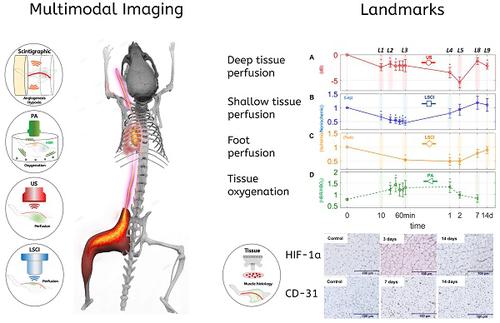
Methods: In order to study the spatiotemporal dynamics of both perfusion and neovascularization in mice subjected to surgically-induced hindlimb ischemia (n= 30), we employed three label-free imaging modalities (a novel high-sensitivity ultrasonic Power Doppler methodology, laser speckle contrast, and photoacoustic imaging), as well as a tandem of radio-labeled molecular probes, 99mTc-NC100692 and 99mTc-BRU-5921 respectively, designed to detect two key modulators of angiogenic activity, αVβ3 and HIF-1α , via scintigraphic imaging.
Results: The multimodal imaging strategy reveals a set of “landmarks”—key physiological and molecular events in the healing process—that can serve as a standardized framework for describing the impact of emerging PAD treatments. These landmarks span the entire process of neovascularization, beginning with the rapid decreases in perfusion and oxygenation associated with ligation surgery, extending through pro-angiogenic changes in gene expression driven by the master regulator HIF-1α , and ultimately leading to complete functional revascularization of the affected tissues.
Conclusions: This study represents an important step in the development of multimodal non-invasive imaging strategies for vascular research; the combined results offer more insight than can be gleaned through any of the individual imaging methods alone. Researchers adopting similar imaging strategies and will be better able to describe changes in the onset, duration, and strength of each of the landmarks of vascular recovery, yielding greater biological insight, and enabling more comprehensive cross-study comparisons. Perhaps most important, this study paves the road for more efficient translation of PAD research; emerging experimental treatments can be more effectively assessed and refined at the preclinical stage, ultimately leading to better next-generation therapies.
中文翻译:
成像血管恢复的地标。
背景:外周动脉疾病(PAD)是世界范围内的主要健康问题。自1990年代后期以来,已经对治疗性血管生成进行了研究,以替代传统的PAD治疗。尽管文献中有大量的临床前阳性结果,但是人类临床试验的结果令人沮丧。该领域面临的挑战之一是缺乏用于验证新治疗方法的时机和措施的标准化。
方法:为了研究小鼠手术引起的后肢缺血(n = 30)的灌注和新血管形成的时空动态,我们采用了三种无标记的成像方式(一种新型的高灵敏度超声功率多普勒方法,激光散斑法)对比度和光声成像),以及放射性标记的分子探针的串联,99米的Tc-NC100692和99米锝BRU-5921分别设计用于检测血管生成活性的两个关键调节剂,α V β 3和HIF-1α ,通过闪烁显像。
结果:多峰成像策略揭示了一组“地标”,即愈合过程中的关键生理和分子事件,可作为描述新兴PAD治疗效果的标准化框架。这些标志性因素涵盖了新血管形成的整个过程,从与结扎手术相关的灌注和氧合迅速减少开始,一直延伸到由主调节因子HIF-1α驱动的基因表达的促血管生成变化,最终导致了完整的功能性血管重建受影响的组织。
结论:本研究代表了用于血管研究的多模式无创成像策略发展的重要一步;组合的结果提供的见解比仅通过任何单个成像方法可以收集的见解要多。研究人员采用类似的成像策略,将能够更好地描述血管恢复的每个标志的发作,持续时间和强度的变化,从而获得更大的生物学见解,并能够进行更全面的跨研究比较。也许最重要的是,这项研究为更有效地翻译PAD研究铺平了道路。可以在临床前阶段更有效地评估和完善新兴的实验治疗方法,最终导致更好的下一代治疗方法。


 京公网安备 11010802027423号