当前位置:
X-MOL 学术
›
PLOS Biol.
›
论文详情
Our official English website, www.x-mol.net, welcomes your
feedback! (Note: you will need to create a separate account there.)
High-resolution 3D imaging and topological mapping of the lymph node conduit system.
PLOS Biology ( IF 7.8 ) Pub Date : 2019-12-19 , DOI: 10.1371/journal.pbio.3000486 Inken D Kelch 1, 2 , Gib Bogle 1, 3 , Gregory B Sands 3 , Anthony R J Phillips 1, 2, 4 , Ian J LeGrice 3, 5 , P Rod Dunbar 1, 2
PLOS Biology ( IF 7.8 ) Pub Date : 2019-12-19 , DOI: 10.1371/journal.pbio.3000486 Inken D Kelch 1, 2 , Gib Bogle 1, 3 , Gregory B Sands 3 , Anthony R J Phillips 1, 2, 4 , Ian J LeGrice 3, 5 , P Rod Dunbar 1, 2
Affiliation
|
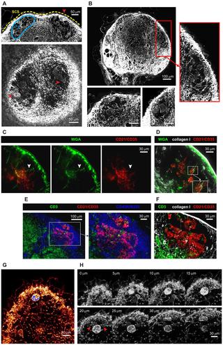
The conduit network is a hallmark of lymph node microanatomy, but lack of suitable imaging technology has prevented comprehensive investigation of its topology. We employed an extended-volume imaging system to capture the conduit network of an entire murine lymph node (comprising over 280,000 segments). The extensive 3D images provide a comprehensive overview of the regions supplied by conduits, including perivascular sleeves and distinctive "follicular reservoirs" within B cell follicles, surrounding follicular dendritic cells. A 3D topology map of conduits within the T-cell zone showed homogeneous branching, but conduit density was significantly higher in the superficial T-cell zone compared with the deep zone, where distances between segments are sufficient for T cells to lose contact with fibroblastic reticular cells. This topological mapping of the conduit anatomy can now aid modeling of its roles in lymph node function, as we demonstrate by simulating T-cell motility in the different T-cell zones.
中文翻译:

淋巴结导管系统的高分辨率3D成像和拓扑图。
导管网络是淋巴结显微解剖的标志,但是缺乏合适的成像技术阻碍了对其拓扑结构的全面研究。我们采用了扩展体积的成像系统来捕获整个鼠淋巴结的导管网络(包括280,000个片段)。广泛的3D图像提供了由导管提供的区域的全面概览,包括血管周套和B细胞滤泡周围,滤泡树突状细胞周围独特的“滤泡储集层”。T细胞区域内导管的3D拓扑图显示了均匀的分支,但与深层区域相比,浅层T细胞区域的导管密度明显更高,在深层区域,节段之间的距离足以使T细胞失去与成纤维网状结构的接触细胞。
更新日期:2019-12-20
中文翻译:
淋巴结导管系统的高分辨率3D成像和拓扑图。
导管网络是淋巴结显微解剖的标志,但是缺乏合适的成像技术阻碍了对其拓扑结构的全面研究。我们采用了扩展体积的成像系统来捕获整个鼠淋巴结的导管网络(包括280,000个片段)。广泛的3D图像提供了由导管提供的区域的全面概览,包括血管周套和B细胞滤泡周围,滤泡树突状细胞周围独特的“滤泡储集层”。T细胞区域内导管的3D拓扑图显示了均匀的分支,但与深层区域相比,浅层T细胞区域的导管密度明显更高,在深层区域,节段之间的距离足以使T细胞失去与成纤维网状结构的接触细胞。


 京公网安备 11010802027423号