当前位置:
X-MOL 学术
›
Lab. Invest.
›
论文详情
Our official English website, www.x-mol.net, welcomes your
feedback! (Note: you will need to create a separate account there.)
Castration-induced stromal remodeling disrupts the reconstituted prostate epithelial structure.
Laboratory Investigation ( IF 5.1 ) Pub Date : 2019-12-19 , DOI: 10.1038/s41374-019-0352-4 Shinya Kajiwara 1 , Kenichiro Ishii 1, 2 , Takeshi Sasaki 1 , Manabu Kato 1 , Kohei Nishikawa 1 , Hideki Kanda 1 , Kiminobu Arima 1 , Masatoshi Watanabe 2 , Yoshiki Sugimura 1
Laboratory Investigation ( IF 5.1 ) Pub Date : 2019-12-19 , DOI: 10.1038/s41374-019-0352-4 Shinya Kajiwara 1 , Kenichiro Ishii 1, 2 , Takeshi Sasaki 1 , Manabu Kato 1 , Kohei Nishikawa 1 , Hideki Kanda 1 , Kiminobu Arima 1 , Masatoshi Watanabe 2 , Yoshiki Sugimura 1
Affiliation
|
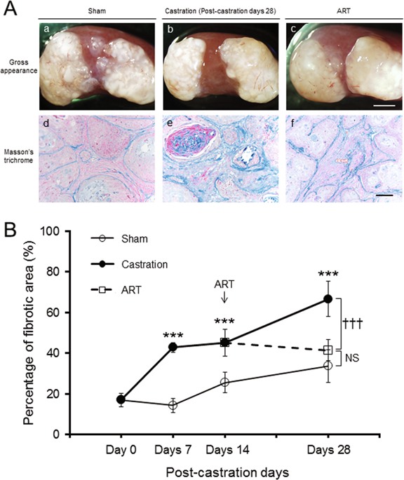
The normal prostate epithelial structure is maintained by homeostatic interactions with smooth muscle cells. However, structural alterations of the stroma are commonly observed in prostatic proliferative diseases, leading to the abnormalities of prostate epithelial structure. A decrease in the androgen level experimentally induces stromal remodeling, i.e., replacement of smooth muscle cells with fibroblasts or myofibroblasts. In this study, we investigated the effects of castration-induced stromal remodeling and subsequent aberrant activation of epithelial-stromal interactions on the reconstituted human prostate-like epithelial structure. We performed in vivo experiments using the human prostate epithelial cell line BPH-1 and fetal rat urogenital sinus mesenchyme to generate heterotypic tissue recombinants that form human prostate-like epithelial structure (i.e., solid- and canalized-epithelial cords). Host mice were castrated at 12 weeks post transplantation (castration) and implanted with a dihydrotestosterone pellet at 14 days post castration (androgen replacement treatment; ART). In the castration group, the percentages of fibrotic area and disrupted prostate epithelial structure without the basement membrane (BM) increased proportionally in a time-dependent manner, but were suppressed by ART. In the castration group, tenascin-C (TNC)-positive fibroblasts were abundant in the stroma surrounding disrupted prostate epithelial structure without the BM. TGF-β1 secretion from BPH-1 cells was increased by co-culturing with human primary cultured prostate fibroblasts. TNC mRNA expression was increased in fibroblasts co-culturing with BPH-1 cells and was suppressed by treatment with a TGF-β RI kinase inhibitor. Moreover, in the castration group, the percentage of p-Smad2-positive cells was significantly higher in the stroma surrounding disrupted prostate epithelial structure without the BM. Our results demonstrate that castration-induced stromal remodeling disrupted the reconstituted human prostate-like epithelial structure and induced the appearance of TNC-positive fibroblasts accompanied by activation of TGF-β signaling. The alteration of prostate stromal structure may be responsible for loss of the BM and epithelial cell polarity.
中文翻译:

去势诱导的基质重塑破坏了重建的前列腺上皮结构。
正常的前列腺上皮结构通过与平滑肌细胞的稳态相互作用来维持。然而,在前列腺增生性疾病中普遍观察到间质的结构改变,导致前列腺上皮结构异常。雄激素水平的降低通过实验诱导基质重塑,即用成纤维细胞或肌成纤维细胞替代平滑肌细胞。在这项研究中,我们研究了去势诱导的间质重塑和随后上皮-间质相互作用的异常激活对重建的人类前列腺样上皮结构的影响。我们使用人前列腺上皮细胞系 BPH-1 和胎鼠泌尿生殖窦间充质进行体内实验,以生成异型组织重组体,形成人前列腺样上皮结构(即固体和管状上皮索)。宿主小鼠在移植后 12 周被阉割(阉割),并在阉割后 14 天植入二氢睾酮颗粒(雄激素替代治疗;ART)。在去势组中,纤维化区域和没有基底膜 (BM) 的前列腺上皮结构破坏的百分比以时间依赖的方式成比例增加,但被 ART 抑制。在去势组中,在没有 BM 的情况下,在破坏的前列腺上皮结构周围的基质中有大量生腱蛋白-C (TNC) 阳性成纤维细胞。通过与人原代培养的前列腺成纤维细胞共培养,BPH-1 细胞的 TGF-β1 分泌增加。TNC mRNA 表达在与 BPH-1 细胞共培养的成纤维细胞中增加,并通过 TGF-β RI 激酶抑制剂处理而受到抑制。此外,在去势组中,在没有 BM 的情况下,在破坏的前列腺上皮结构周围的基质中 p-Smad2 阳性细胞的百分比明显更高。我们的结果表明,去势诱导的基质重塑破坏了重建的人前列腺样上皮结构,并诱导了 TNC 阳性成纤维细胞的出现,同时激活了 TGF-β 信号传导。前列腺间质结构的改变可能是 BM 和上皮细胞极性丧失的原因。
更新日期:2019-12-20
中文翻译:
去势诱导的基质重塑破坏了重建的前列腺上皮结构。
正常的前列腺上皮结构通过与平滑肌细胞的稳态相互作用来维持。然而,在前列腺增生性疾病中普遍观察到间质的结构改变,导致前列腺上皮结构异常。雄激素水平的降低通过实验诱导基质重塑,即用成纤维细胞或肌成纤维细胞替代平滑肌细胞。在这项研究中,我们研究了去势诱导的间质重塑和随后上皮-间质相互作用的异常激活对重建的人类前列腺样上皮结构的影响。我们使用人前列腺上皮细胞系 BPH-1 和胎鼠泌尿生殖窦间充质进行体内实验,以生成异型组织重组体,形成人前列腺样上皮结构(即固体和管状上皮索)。宿主小鼠在移植后 12 周被阉割(阉割),并在阉割后 14 天植入二氢睾酮颗粒(雄激素替代治疗;ART)。在去势组中,纤维化区域和没有基底膜 (BM) 的前列腺上皮结构破坏的百分比以时间依赖的方式成比例增加,但被 ART 抑制。在去势组中,在没有 BM 的情况下,在破坏的前列腺上皮结构周围的基质中有大量生腱蛋白-C (TNC) 阳性成纤维细胞。通过与人原代培养的前列腺成纤维细胞共培养,BPH-1 细胞的 TGF-β1 分泌增加。TNC mRNA 表达在与 BPH-1 细胞共培养的成纤维细胞中增加,并通过 TGF-β RI 激酶抑制剂处理而受到抑制。此外,在去势组中,在没有 BM 的情况下,在破坏的前列腺上皮结构周围的基质中 p-Smad2 阳性细胞的百分比明显更高。我们的结果表明,去势诱导的基质重塑破坏了重建的人前列腺样上皮结构,并诱导了 TNC 阳性成纤维细胞的出现,同时激活了 TGF-β 信号传导。前列腺间质结构的改变可能是 BM 和上皮细胞极性丧失的原因。


 京公网安备 11010802027423号