Our official English website, www.x-mol.net, welcomes your
feedback! (Note: you will need to create a separate account there.)
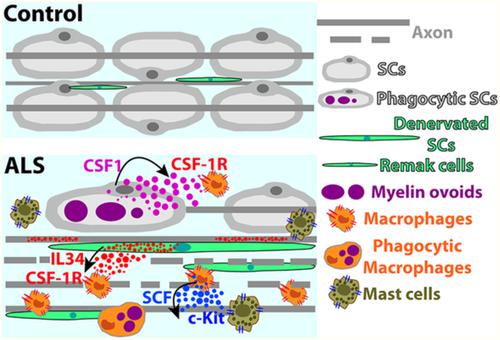
Schwann cells orchestrate peripheral nerve inflammation through the expression of CSF1, IL-34, and SCF in amyotrophic lateral sclerosis.
Glia ( IF 5.4 ) Pub Date : 2019-12-20 , DOI: 10.1002/glia.23768 Emiliano Trias 1 , Mariángeles Kovacs 1 , Peter H King 2, 3 , Ying Si 2, 3 , Yuri Kwon 2 , Valentina Varela 1 , Sofía Ibarburu 1 , Ivan C Moura 4, 5, 6, 7, 8, 9 , Olivier Hermine 4, 5, 6, 7, 8, 9, 10, 11, 12 , Joseph S Beckman 13 , Luis Barbeito 1
Glia ( IF 5.4 ) Pub Date : 2019-12-20 , DOI: 10.1002/glia.23768 Emiliano Trias 1 , Mariángeles Kovacs 1 , Peter H King 2, 3 , Ying Si 2, 3 , Yuri Kwon 2 , Valentina Varela 1 , Sofía Ibarburu 1 , Ivan C Moura 4, 5, 6, 7, 8, 9 , Olivier Hermine 4, 5, 6, 7, 8, 9, 10, 11, 12 , Joseph S Beckman 13 , Luis Barbeito 1
Affiliation
|
Distal axonopathy is a recognized pathological feature of amyotrophic lateral sclerosis (ALS). In the peripheral nerves of ALS patients, motor axon loss elicits a Wallerian-like degeneration characterized by denervated Schwann cells (SCs) together with immune cell infiltration. However, the pathogenic significance of denervated SCs accumulating following impaired axonal growth in ALS remains unclear. Here, we analyze SC phenotypes in sciatic nerves of ALS patients and paralytic SOD1G93A rats, and identify remarkably similar and specific reactive SC phenotypes based on the pattern of S100β, GFAP, isolectin and/or p75NTR immunoreactivity. Different subsets of reactive SCs expressed colony-stimulating factor-1 (CSF1) and Interleukin-34 (IL-34) and closely interacted with numerous endoneurial CSF-1R-expressing monocyte/macrophages, suggesting a paracrine mechanism of myeloid cell expansion and activation. SCs bearing phagocytic phenotypes as well as endoneurial macrophages expressed stem cell factor (SCF), a trophic factor that attracts and activates mast cells through the c-Kit receptor. Notably, a subpopulation of Ki67+ SCs expressed c-Kit in the sciatic nerves of SOD1G93A rats, suggesting a signaling pathway that fuels SC proliferation in ALS. c-Kit+ mast cells were also abundant in the sciatic nerve from ALS donors but not in controls. Pharmacological inhibition of CSF-1R and c-Kit with masitinib in SOD1G93A rats potently reduced SC reactivity and immune cell infiltration in the sciatic nerve and ventral roots, suggesting a mechanism by which the drug ameliorates peripheral nerve pathology. These findings provide strong evidence for a previously unknown inflammatory mechanism triggered by SCs in ALS peripheral nerves that has broad application in developing novel therapies.
中文翻译:

雪旺氏细胞通过在肌萎缩侧索硬化中表达 CSF1、IL-34 和 SCF 来协调外周神经炎症。
远端轴突病是肌萎缩侧索硬化 (ALS) 公认的病理特征。在 ALS 患者的外周神经中,运动轴突缺失会引起沃勒样变性,其特征是去神经支配的雪旺细胞 (SCs) 以及免疫细胞浸润。然而,在 ALS 中轴突生长受损后积累的去神经支配的 SCs 的致病意义仍不清楚。在这里,我们分析了 ALS 患者和麻痹性 SOD1G93A 大鼠坐骨神经中的 SC 表型,并根据 S100β、GFAP、同种凝集素和/或 p75NTR 免疫反应性模式确定了非常相似和特异性的反应性 SC 表型。反应性 SCs 的不同亚群表达集落刺激因子-1 (CSF1) 和白细胞介素-34 (IL-34),并与许多表达神经内膜 CSF-1R 的单核细胞/巨噬细胞密切相互作用,表明骨髓细胞扩增和激活的旁分泌机制。具有吞噬细胞表型的 SC 以及神经内膜巨噬细胞表达干细胞因子 (SCF),这是一种通过 c-Kit 受体吸引和激活肥大细胞的营养因子。值得注意的是,一个 Ki67+ SCs 亚群在 SOD1G93A 大鼠的坐骨神经中表达了 c-Kit,这表明信号通路促进了 ALS 中的 SC 增殖。c-Kit+ 肥大细胞在 ALS 供体的坐骨神经中也很丰富,但在对照组中则不然。在 SOD1G93A 大鼠中使用马赛替尼对 CSF-1R 和 c-Kit 的药理学抑制有效降低了坐骨神经和腹神经根的 SC 反应性和免疫细胞浸润,表明该药物改善周围神经病理的机制。
更新日期:2019-12-20
中文翻译:
雪旺氏细胞通过在肌萎缩侧索硬化中表达 CSF1、IL-34 和 SCF 来协调外周神经炎症。
远端轴突病是肌萎缩侧索硬化 (ALS) 公认的病理特征。在 ALS 患者的外周神经中,运动轴突缺失会引起沃勒样变性,其特征是去神经支配的雪旺细胞 (SCs) 以及免疫细胞浸润。然而,在 ALS 中轴突生长受损后积累的去神经支配的 SCs 的致病意义仍不清楚。在这里,我们分析了 ALS 患者和麻痹性 SOD1G93A 大鼠坐骨神经中的 SC 表型,并根据 S100β、GFAP、同种凝集素和/或 p75NTR 免疫反应性模式确定了非常相似和特异性的反应性 SC 表型。反应性 SCs 的不同亚群表达集落刺激因子-1 (CSF1) 和白细胞介素-34 (IL-34),并与许多表达神经内膜 CSF-1R 的单核细胞/巨噬细胞密切相互作用,表明骨髓细胞扩增和激活的旁分泌机制。具有吞噬细胞表型的 SC 以及神经内膜巨噬细胞表达干细胞因子 (SCF),这是一种通过 c-Kit 受体吸引和激活肥大细胞的营养因子。值得注意的是,一个 Ki67+ SCs 亚群在 SOD1G93A 大鼠的坐骨神经中表达了 c-Kit,这表明信号通路促进了 ALS 中的 SC 增殖。c-Kit+ 肥大细胞在 ALS 供体的坐骨神经中也很丰富,但在对照组中则不然。在 SOD1G93A 大鼠中使用马赛替尼对 CSF-1R 和 c-Kit 的药理学抑制有效降低了坐骨神经和腹神经根的 SC 反应性和免疫细胞浸润,表明该药物改善周围神经病理的机制。


 京公网安备 11010802027423号