当前位置:
X-MOL 学术
›
Macromol. Rapid Commun.
›
论文详情
Our official English website, www.x-mol.net, welcomes your
feedback! (Note: you will need to create a separate account there.)
Characterizing Cross-Linking Within Polymeric Biomaterials in the SEM by Secondary Electron Hyperspectral Imaging.
Macromolecular Rapid Communications ( IF 4.2 ) Pub Date : 2019-12-20 , DOI: 10.1002/marc.201900484 Nicholas Farr 1 , Samand Pashneh-Tala 1 , Nicola Stehling 1 , Frederik Claeyssens 1 , Nicola Green 1, 2 , Cornelia Rodenburg 1
Macromolecular Rapid Communications ( IF 4.2 ) Pub Date : 2019-12-20 , DOI: 10.1002/marc.201900484 Nicholas Farr 1 , Samand Pashneh-Tala 1 , Nicola Stehling 1 , Frederik Claeyssens 1 , Nicola Green 1, 2 , Cornelia Rodenburg 1
Affiliation
|
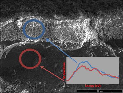
A novel capability built upon secondary electron (SE) spectroscopy provides an enhanced cross-linking characterization toolset for polymeric biomaterials, with cross-linking density and variation captured at a multiscale level. The potential of SE spectroscopy for material characterization has been investigated since 1947. The absence of suitable instrumentation and signal processing proved insurmountable barriers to applying SE spectroscopy to biomaterials, and consequently, capturing SE spectra containing cross-linking information is a new concept. To date, cross-linking extent is inferred from analytical techniques such as nuclear magnetic resonance (NMR), differential scanning calorimetry, and Raman spectroscopy (RS). NMR provides extremely localized information on the atomic scale and molecular scale, while RS information volume is on the microscale. Other methods for the indirect study of cross-linking are bulk mechanical averaging methods, such as tensile and compression modulus testing. However, these established averaging methods for the estimation of polymer cross-linking density are incomplete because they fail to provide information of spatial distributions within the biomaterial morphology across all relevant length scales. The efficacy of the SE spectroscopy capability is demonstrated in this paper by the analysis of poly(glycerol sebacate)-methacrylate (PGS-M) at different degrees of methacrylation delivering new insights into PGS-M morphology.
中文翻译:

通过二次电子高光谱成像表征SEM中高分子生物材料内的交联。
基于二次电子(SE)光谱学的新型功能为聚合物生物材料提供了增强的交联表征工具集,具有在多尺度水平上捕获的交联密度和变化。自1947年以来,人们一直在研究SE光谱在材料表征中的潜力。缺乏合适的仪器和信号处理证明了将SE光谱应用于生物材料的不可克服的障碍,因此,捕获包含交联信息的SE光谱是一个新概念。迄今为止,可以从诸如核磁共振(NMR),差示扫描量热法和拉曼光谱法(RS)之类的分析技术推断出交联程度。NMR可提供有关原子级和分子级的极其局部的信息,而RS信息量是微尺度的。间接研究交联的其他方法是本体机械平均法,例如拉伸和压缩模量测试。但是,这些建立的用于估计聚合物交联密度的平均方法是不完整的,因为它们无法提供所有相关长度范围内生物材料形态内空间分布的信息。通过在不同甲基丙烯酸化程度下对聚癸二酸甘油酯-甲基丙烯酸酯(PGS-M)进行分析,证明了SE光谱学功能的功效,从而为PGS-M形态提供了新的见识。这些建立的用于估计聚合物交联密度的平均方法是不完整的,因为它们无法提供所有相关长度范围内生物材料形态内空间分布的信息。通过对聚甲基丙烯酸癸二酯-甲基丙烯酸酯(PGS-M)在不同甲基丙烯酸化程度下的分析,证明了SE光谱学功能的有效性,从而为PGS-M形态学提供了新的见识。这些建立的用于估计聚合物交联密度的平均方法是不完整的,因为它们无法提供所有相关长度范围内生物材料形态内空间分布的信息。通过对聚甲基丙烯酸癸二酯-甲基丙烯酸酯(PGS-M)在不同甲基丙烯酸化程度下的分析,证明了SE光谱学功能的有效性,从而为PGS-M形态学提供了新的见识。
更新日期:2019-12-20
中文翻译:
通过二次电子高光谱成像表征SEM中高分子生物材料内的交联。
基于二次电子(SE)光谱学的新型功能为聚合物生物材料提供了增强的交联表征工具集,具有在多尺度水平上捕获的交联密度和变化。自1947年以来,人们一直在研究SE光谱在材料表征中的潜力。缺乏合适的仪器和信号处理证明了将SE光谱应用于生物材料的不可克服的障碍,因此,捕获包含交联信息的SE光谱是一个新概念。迄今为止,可以从诸如核磁共振(NMR),差示扫描量热法和拉曼光谱法(RS)之类的分析技术推断出交联程度。NMR可提供有关原子级和分子级的极其局部的信息,而RS信息量是微尺度的。间接研究交联的其他方法是本体机械平均法,例如拉伸和压缩模量测试。但是,这些建立的用于估计聚合物交联密度的平均方法是不完整的,因为它们无法提供所有相关长度范围内生物材料形态内空间分布的信息。通过在不同甲基丙烯酸化程度下对聚癸二酸甘油酯-甲基丙烯酸酯(PGS-M)进行分析,证明了SE光谱学功能的功效,从而为PGS-M形态提供了新的见识。这些建立的用于估计聚合物交联密度的平均方法是不完整的,因为它们无法提供所有相关长度范围内生物材料形态内空间分布的信息。通过对聚甲基丙烯酸癸二酯-甲基丙烯酸酯(PGS-M)在不同甲基丙烯酸化程度下的分析,证明了SE光谱学功能的有效性,从而为PGS-M形态学提供了新的见识。这些建立的用于估计聚合物交联密度的平均方法是不完整的,因为它们无法提供所有相关长度范围内生物材料形态内空间分布的信息。通过对聚甲基丙烯酸癸二酯-甲基丙烯酸酯(PGS-M)在不同甲基丙烯酸化程度下的分析,证明了SE光谱学功能的有效性,从而为PGS-M形态学提供了新的见识。


 京公网安备 11010802027423号