当前位置:
X-MOL 学术
›
Acta Biomater.
›
论文详情
Our official English website, www.x-mol.net, welcomes your
feedback! (Note: you will need to create a separate account there.)
Evaluation of a conducting elastomeric composite material for intramuscular electrode application.
Acta Biomaterialia ( IF 9.4 ) Pub Date : 2019-12-18 , DOI: 10.1016/j.actbio.2019.12.021 X Sally Zheng 1 , Azante Y Griffith 1 , Emily Chang 2 , Michael J Looker 2 , Lee E Fisher 3 , Brady Clapsaddle 2 , X Tracy Cui 4
Acta Biomaterialia ( IF 9.4 ) Pub Date : 2019-12-18 , DOI: 10.1016/j.actbio.2019.12.021 X Sally Zheng 1 , Azante Y Griffith 1 , Emily Chang 2 , Michael J Looker 2 , Lee E Fisher 3 , Brady Clapsaddle 2 , X Tracy Cui 4
Affiliation
|
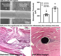
Electrical stimulation of the muscle has been proven efficacious in preventing atrophy and/or reanimating paralyzed muscles. Intramuscular electrodes made from metals have significantly higher Young's Moduli than the muscle tissues, which has the potential to cause chronic inflammation and decrease device performance. Here, we present an intramuscular electrode made from an elastomeric conducting polymer composite consisting of PEDOT-PEG copolymer, silicone and carbon nanotubes (CNT) with fluorosilicone insulation. The electrode wire has a Young's modulus of 804 (±99) kPa, which better mimics the muscle tissue modulus than conventional stainless steel (SS) electrodes. Additionally, the non-metallic composition enables metal-artifact free CT and MR imaging. These soft wire (SW) electrodes present comparable electrical impedance to SS electrodes of similar geometric surface area, activate muscle at a lower threshold, and maintain stable electrical properties in vivo up to 4 weeks. Histologically, the SW electrodes elicited significantly less fibrotic encapsulation and less IBA-1 positive macrophage accumulation than the SS electrodes at one and three months. Further phenotyping the macrophages with the iNOS (pro-inflammatory) and ARG-1 (pro-healing) markers revealed significantly less presence of pro-inflammatory macrophage around SW implants at one month. By three months, there was a significant increase in pro-healing macrophages (ARG-1) around the SW implants but not around the SS implants. Furthermore, a larger number of AchR clusters closer to SW implants were found at both time points compared to SS implants. These results suggest that a softer implant encourages a more intimate and healthier electrode-tissue interface. STATEMENT OF SIGNIFICANCE: Intramuscular electrodes made from metals have significantly higher Young's Moduli than the muscle tissues, which has the potential to cause chronic inflammation and decrease device performance. Here, we present an intramuscular electrode made from an elastomeric conducting polymer composite consisting of PEDOT-PEG copolymer, silicone and carbon nanotubes with fluorosilicone insulation. This elastomeric composite results in an electrode wire with a Young's modulus mimicking that of the muscle tissue, which elicits significantly less foreign body response compared to stainless steel wires. The lack of metal in this composite also enables metal-artifact free MRI and CT imaging.
中文翻译:

肌内电极应用的导电弹性复合材料的评估。
肌肉的电刺激已被证明在预防萎缩和/或使瘫痪的肌肉恢复活力方面是有效的。由金属制成的肌内电极比肌肉组织具有更高的杨氏模量,这有可能引起慢性炎症并降低器械性能。在这里,我们介绍了一种肌内电极,该肌内电极由弹性体导电聚合物复合材料制成,该复合材料由PEDOT-PEG共聚物,硅树脂和具有氟硅氧烷绝缘层的碳纳米管(CNT)组成。电极丝的杨氏模量为804(±99)kPa,比传统的不锈钢(SS)电极更好地模仿了肌肉组织的模量。另外,非金属组合物使得无金属伪像的CT和MR成像成为可能。这些软线(SW)电极具有与具有相似几何表面积的SS电极相当的电阻抗,可以在较低的阈值处激活肌肉,并在长达4周的时间内保持体内稳定的电性能。从组织学上讲,与SS电极相比,SW电极在1个月和3个月时引起的纤维化包囊和IBA-1阳性巨噬细胞的积聚明显更少。用iNOS(促炎性)和ARG-1(促愈合)标记对巨噬细胞进行进一步的表型分析显示,在一个月内,SW植入物周围促炎性巨噬细胞的存在显着减少。到三个月时,SW植入物周围的愈合前巨噬细胞(ARG-1)显着增加,而SS植入物周围的则没有。此外,与SS植入物相比,在两个时间点都发现有更多的AchR簇更接近SW植入物。这些结果表明,较软的植入物可促进更紧密和更健康的电极-组织界面。意义声明:由金属制成的肌内电极比肌肉组织具有更高的杨氏模量,这有可能引起慢性炎症并降低器械性能。在这里,我们介绍了一种肌内电极,该肌内电极由弹性体导电聚合物复合材料制成,该复合材料由PEDOT-PEG共聚物,有机硅和具有氟硅氧烷绝缘层的碳纳米管组成。这种弹性复合材料产生的电极线的杨氏模量类似于肌肉组织的杨氏模量,与不锈钢线相比,它引起的异物响应明显更少。
更新日期:2019-12-19
中文翻译:
肌内电极应用的导电弹性复合材料的评估。
肌肉的电刺激已被证明在预防萎缩和/或使瘫痪的肌肉恢复活力方面是有效的。由金属制成的肌内电极比肌肉组织具有更高的杨氏模量,这有可能引起慢性炎症并降低器械性能。在这里,我们介绍了一种肌内电极,该肌内电极由弹性体导电聚合物复合材料制成,该复合材料由PEDOT-PEG共聚物,硅树脂和具有氟硅氧烷绝缘层的碳纳米管(CNT)组成。电极丝的杨氏模量为804(±99)kPa,比传统的不锈钢(SS)电极更好地模仿了肌肉组织的模量。另外,非金属组合物使得无金属伪像的CT和MR成像成为可能。这些软线(SW)电极具有与具有相似几何表面积的SS电极相当的电阻抗,可以在较低的阈值处激活肌肉,并在长达4周的时间内保持体内稳定的电性能。从组织学上讲,与SS电极相比,SW电极在1个月和3个月时引起的纤维化包囊和IBA-1阳性巨噬细胞的积聚明显更少。用iNOS(促炎性)和ARG-1(促愈合)标记对巨噬细胞进行进一步的表型分析显示,在一个月内,SW植入物周围促炎性巨噬细胞的存在显着减少。到三个月时,SW植入物周围的愈合前巨噬细胞(ARG-1)显着增加,而SS植入物周围的则没有。此外,与SS植入物相比,在两个时间点都发现有更多的AchR簇更接近SW植入物。这些结果表明,较软的植入物可促进更紧密和更健康的电极-组织界面。意义声明:由金属制成的肌内电极比肌肉组织具有更高的杨氏模量,这有可能引起慢性炎症并降低器械性能。在这里,我们介绍了一种肌内电极,该肌内电极由弹性体导电聚合物复合材料制成,该复合材料由PEDOT-PEG共聚物,有机硅和具有氟硅氧烷绝缘层的碳纳米管组成。这种弹性复合材料产生的电极线的杨氏模量类似于肌肉组织的杨氏模量,与不锈钢线相比,它引起的异物响应明显更少。


 京公网安备 11010802027423号