当前位置:
X-MOL 学术
›
Cell Death Discov.
›
论文详情
Our official English website, www.x-mol.net, welcomes your
feedback! (Note: you will need to create a separate account there.)
Physical disruption of intervertebral disc promotes cell clustering and a degenerative phenotype.
Cell Death Discovery ( IF 6.1 ) Pub Date : 2019-12-17 , DOI: 10.1038/s41420-019-0233-z Polly Lama 1 , Harry Claireaux 2 , Luke Flower 3 , Ian J Harding 4 , Trish Dolan 5 , Christine L Le Maitre 6 , Michael A Adams 5
Cell Death Discovery ( IF 6.1 ) Pub Date : 2019-12-17 , DOI: 10.1038/s41420-019-0233-z Polly Lama 1 , Harry Claireaux 2 , Luke Flower 3 , Ian J Harding 4 , Trish Dolan 5 , Christine L Le Maitre 6 , Michael A Adams 5
Affiliation
|
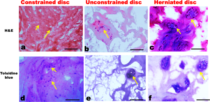
To test the hypothesis that physical disruption of an intervertebral disc disturbs cell-matrix binding, leading to cell clustering and increased expression of matrix degrading enzymes that contribute towards degenerative disc cell phenotype. Lumbar disc tissue was removed at surgery from 21 patients with disc herniation, 11 with disc degeneration, and 8 with adolescent scoliosis. 5 μm sections were examined with histology, and 30-µm sections by confocal microscopy. Antibodies were used against integrin α5beta1, matrix metalloproteinases (MMP) 1, MMP-3, caspase 3, and denatured collagen types I and II. Spatial associations were sought between cell clustering and various degenerative features. An additional, 11 non-herniated human discs were used to examine causality: half of each specimen was cultured in a manner that allowed free 'unconstrained' swelling (similar to a herniated disc in vivo), while the other half was cultured within a perspex ring that allowed 'constrained' swelling. Changes were monitored over 36 h using live-cell imaging. 1,9-Di-methyl methylene blue (DMMB) assay for glycosaminoglycan loss was carried out from tissue medium. Partially constrained specimens showed little swelling or cell movement in vitro. In contrast, unconstrained swelling significantly increased matrix distortion, glycosaminoglycan loss, exposure of integrin binding sites, expression of MMPs 1 and 3, and collagen denaturation. In the association studies, herniated disc specimens showed changes that resembled unconstrained swelling in vitro. In addition, they exhibited increased cell clustering, apoptosis, MMP expression, and collagen denaturation compared to 'control' discs. Results support our hypothesis. Further confirmation will require longitudinal animal experiments.
中文翻译:

椎间盘的物理破坏促进细胞聚集和变性表型。
为了检验假说,椎间盘的物理破坏会干扰细胞-基质结合,从而导致细胞聚集和基质降解酶表达的增加,从而导致椎间盘退变的细胞表型。手术时从21例椎间盘突出症患者,11例椎间盘退变患者和8例青少年脊柱侧弯患者中取出腰椎间盘组织。用组织学检查5μm切片,通过共聚焦显微镜检查30μm切片。抗体用于整合素α5beta1,基质金属蛋白酶(MMP)1,MMP-3,胱天蛋白酶3和变性的I型和II型胶原蛋白。在细胞聚类和各种退化特征之间寻求空间关联。另外,使用了11个非突出的人类椎间盘来检查因果关系:每个标本的一半都以允许自由“移动”的方式进行培养。不受限制的肿胀(类似于体内椎间盘突出),而另一半则在允许“受限制的”肿胀的有机玻璃环中培养。使用活细胞成像监测变化超过36小时。从组织培养基中进行1,9-二甲基亚甲基蓝(DMMB)糖胺聚糖损失的测定。部分受约束的标本在体外几乎没有肿胀或细胞运动。相反,不受约束的膨胀显着增加了基质变形,糖胺聚糖损失,整联蛋白结合位点的暴露,MMP 1和3的表达以及胶原变性。在关联研究中,椎间盘突出的标本显示出类似于不受约束的体外肿胀的变化。此外,与“对照”相比,它们表现出增加的细胞簇集,凋亡,MMP表达和胶原变性 光盘。结果支持我们的假设。进一步的确认将需要纵向动物实验。
更新日期:2019-12-17
中文翻译:
椎间盘的物理破坏促进细胞聚集和变性表型。
为了检验假说,椎间盘的物理破坏会干扰细胞-基质结合,从而导致细胞聚集和基质降解酶表达的增加,从而导致椎间盘退变的细胞表型。手术时从21例椎间盘突出症患者,11例椎间盘退变患者和8例青少年脊柱侧弯患者中取出腰椎间盘组织。用组织学检查5μm切片,通过共聚焦显微镜检查30μm切片。抗体用于整合素α5beta1,基质金属蛋白酶(MMP)1,MMP-3,胱天蛋白酶3和变性的I型和II型胶原蛋白。在细胞聚类和各种退化特征之间寻求空间关联。另外,使用了11个非突出的人类椎间盘来检查因果关系:每个标本的一半都以允许自由“移动”的方式进行培养。不受限制的肿胀(类似于体内椎间盘突出),而另一半则在允许“受限制的”肿胀的有机玻璃环中培养。使用活细胞成像监测变化超过36小时。从组织培养基中进行1,9-二甲基亚甲基蓝(DMMB)糖胺聚糖损失的测定。部分受约束的标本在体外几乎没有肿胀或细胞运动。相反,不受约束的膨胀显着增加了基质变形,糖胺聚糖损失,整联蛋白结合位点的暴露,MMP 1和3的表达以及胶原变性。在关联研究中,椎间盘突出的标本显示出类似于不受约束的体外肿胀的变化。此外,与“对照”相比,它们表现出增加的细胞簇集,凋亡,MMP表达和胶原变性 光盘。结果支持我们的假设。进一步的确认将需要纵向动物实验。


 京公网安备 11010802027423号