当前位置:
X-MOL 学术
›
npj Precis. Oncol.
›
论文详情
Our official English website, www.x-mol.net, welcomes your
feedback! (Note: you will need to create a separate account there.)
Real-time intraoperative diagnosis by deep neural network driven multiphoton virtual histology.
npj Precision Oncology ( IF 6.8 ) Pub Date : 2019-12-17 , DOI: 10.1038/s41698-019-0104-3 Sixian You 1, 2 , Yi Sun 1, 3 , Lin Yang 4 , Jaena Park 1, 2 , Haohua Tu 1 , Marina Marjanovic 1, 2, 5 , Saurabh Sinha 5, 6 , Stephen A Boppart 1, 2, 3, 5
npj Precision Oncology ( IF 6.8 ) Pub Date : 2019-12-17 , DOI: 10.1038/s41698-019-0104-3 Sixian You 1, 2 , Yi Sun 1, 3 , Lin Yang 4 , Jaena Park 1, 2 , Haohua Tu 1 , Marina Marjanovic 1, 2, 5 , Saurabh Sinha 5, 6 , Stephen A Boppart 1, 2, 3, 5
Affiliation
|
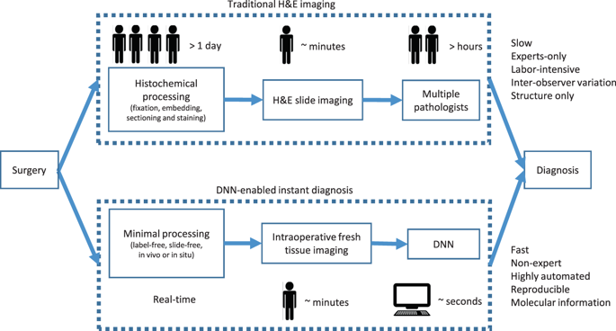
Recent advances in label-free virtual histology promise a new era for real-time molecular diagnosis in the operating room and during biopsy procedures. To take full advantage of the rich, multidimensional information provided by these technologies, reproducible and reliable computational tools that could facilitate the diagnosis are in great demand. In this study, we developed a deep-learning-based framework to recognize cancer versus normal human breast tissue from real-time label-free virtual histology images, with a tile-level AUC (area under receiver operating curve) of 95% and slide-level AUC of 100% on unseen samples. Furthermore, models trained on a high-quality laboratory-generated dataset can generalize to independent datasets acquired from a portable intraoperative version of the imaging technology with a physics-based adapted design. Classification activation maps and final feature visualization revealed discriminative patterns, such as tumor cells and tumor-associated vesicles, that are highly associated with cancer status. These results demonstrate that through the combination of real-time virtual histopathology and a deep-learning framework, accurate real-time diagnosis could be achieved in point-of-procedure clinical applications.
中文翻译:

通过深度神经网络驱动的多光子虚拟组织学进行实时术中诊断。
无标签虚拟组织学的最新进展为手术室和活检过程中的实时分子诊断开辟了新纪元。为了充分利用这些技术提供的丰富,多维的信息,迫切需要可帮助诊断的可再现且可靠的计算工具。在这项研究中,我们开发了一种基于深度学习的框架,可从实时的无标签虚拟组织学图像中识别癌症与正常人的乳房组织,其中图块级别的AUC(接收者操作曲线下的面积)为95%,并且滑动看不见的样本的100%级别AUC。此外,在高质量的实验室生成的数据集上训练的模型可以推广到通过基于物理的适应性设计从成像技术的便携式术中版本获取的独立数据集。分类激活图和最终特征可视化揭示了与癌症状态高度相关的判别模式,例如肿瘤细胞和与肿瘤相关的囊泡。这些结果表明,通过将实时虚拟组织病理学和深度学习框架相结合,可以在过程点临床应用中实现准确的实时诊断。
更新日期:2019-12-17
中文翻译:
通过深度神经网络驱动的多光子虚拟组织学进行实时术中诊断。
无标签虚拟组织学的最新进展为手术室和活检过程中的实时分子诊断开辟了新纪元。为了充分利用这些技术提供的丰富,多维的信息,迫切需要可帮助诊断的可再现且可靠的计算工具。在这项研究中,我们开发了一种基于深度学习的框架,可从实时的无标签虚拟组织学图像中识别癌症与正常人的乳房组织,其中图块级别的AUC(接收者操作曲线下的面积)为95%,并且滑动看不见的样本的100%级别AUC。此外,在高质量的实验室生成的数据集上训练的模型可以推广到通过基于物理的适应性设计从成像技术的便携式术中版本获取的独立数据集。分类激活图和最终特征可视化揭示了与癌症状态高度相关的判别模式,例如肿瘤细胞和与肿瘤相关的囊泡。这些结果表明,通过将实时虚拟组织病理学和深度学习框架相结合,可以在过程点临床应用中实现准确的实时诊断。


 京公网安备 11010802027423号