Our official English website, www.x-mol.net, welcomes your
feedback! (Note: you will need to create a separate account there.)
Addressing the presence of biogenic selenium nanoparticles in yeast cells: analytical strategies based on ICP-TQ-MS.
Analyst ( IF 3.6 ) Pub Date : 2019-12-23 , DOI: 10.1039/c9an01565e R Álvarez-Fernández García 1 , M Corte-Rodríguez , M Macke , K L LeBlanc , Z Mester , M Montes-Bayón , J Bettmer
Analyst ( IF 3.6 ) Pub Date : 2019-12-23 , DOI: 10.1039/c9an01565e R Álvarez-Fernández García 1 , M Corte-Rodríguez , M Macke , K L LeBlanc , Z Mester , M Montes-Bayón , J Bettmer
Affiliation
|
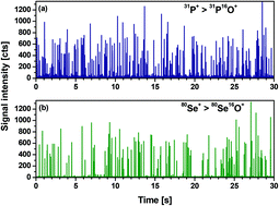
Several organisms have demonstrated the ability of synthesising biogenic selenium-containing nanoparticles. Such particles from biological sources have attracted great attention due to several proven activities as antioxidants or antimicrobial agents. However, little is known in terms of size (distribution), shapes, chemical composition and number/amount/concentration of these particles. Therefore, in this work, we proposed the use of complementary analytical strategies that enabled the detection and characterization of selenium-containing nanoparticles in selenized yeast (Saccharomyces cerevisiae). The first strategy to address the intracellular presence of Se within yeast cells, involves the use of single cell ICP-TQ-MS (inductively coupled plasma-mass spectrometry). For this aim, selenium and phosphorous (as constitutive element) were measured as oxides (80Se16O+ and 31P16O+, resp.) in the triple-quadrupole mode. Then, a simple and fast cell lysis by mechanical disruption is conducted (approx. 30 min) in order to prove the presence of selenium-containing nanoparticles (SeNPs). The lysate is analysed by single particle ICP-TQ-MS and, complementarily, by liquid chromatography coupled to ICP-TQ-MS to cover a wider range of particle sizes. One of the samples revealed the presence of dispersed SeNPs with sizes between a few nm and up to 250 nm also confirmed by transmission electron microscopy (TEM) in the form of elemental selenium. The analysis of the certified reference material SELM-1 showed the presence of spherical SeNPs of 4 to 7 nm diameter. These biogenic particles, at least partially, were made of elemental selenium as well. The whole study reveals the excellent capabilities of "single" event ICP-MS methodologies in combination with HPLC-based strategies for a complete characterization of nanoparticulated material in biological samples.
中文翻译:

解决酵母细胞中生物硒纳米颗粒的存在:基于ICP-TQ-MS的分析策略。
几种生物体已证明具有合成生物来源的含硒纳米粒子的能力。由于数种已证明的抗氧化剂或抗菌剂活性,因此来自生物来源的此类颗粒引起了极大的关注。然而,就这些颗粒的尺寸(分布),形状,化学组成和数量/数量/浓度而言知之甚少。因此,在这项工作中,我们提出了使用互补分析策略的方法,该策略能够检测和表征硒化酵母(Saccharomyces cerevisiae)中的含硒纳米颗粒。解决酵母细胞内硒在细胞内存在的第一种策略涉及使用单细胞ICP-TQ-MS(电感耦合等离子体质谱法)。为了这个目的,硒和磷(作为构成元素)以三重四极杆模式作为氧化物(分别为80Se16O +和31P16O +)进行测量。然后,通过机械破坏进行简单,快速的细胞裂解(约30分钟),以证明存在含硒的纳米颗粒(SeNPs)。裂解物通过单颗粒ICP-TQ-MS进行分析,补充地通过与ICP-TQ-MS联用的液相色谱法进行分析,以涵盖更大范围的粒径。其中一个样品表明存在分散的SeNPs,其尺寸在几nm至250 nm之间,并通过透射电子显微镜(TEM)以元素硒的形式得到证实。经认证的参考材料SELM-1的分析表明,存在直径为4至7 nm的球形SeNP。这些生物微粒至少部分地 也是由元素硒制成的。整个研究揭示了“单一”事件ICP-MS方法学与基于HPLC的策略相结合,可对生物样品中的纳米颗粒材料进行完整表征的卓越能力。
更新日期:2020-02-17
中文翻译:
解决酵母细胞中生物硒纳米颗粒的存在:基于ICP-TQ-MS的分析策略。
几种生物体已证明具有合成生物来源的含硒纳米粒子的能力。由于数种已证明的抗氧化剂或抗菌剂活性,因此来自生物来源的此类颗粒引起了极大的关注。然而,就这些颗粒的尺寸(分布),形状,化学组成和数量/数量/浓度而言知之甚少。因此,在这项工作中,我们提出了使用互补分析策略的方法,该策略能够检测和表征硒化酵母(Saccharomyces cerevisiae)中的含硒纳米颗粒。解决酵母细胞内硒在细胞内存在的第一种策略涉及使用单细胞ICP-TQ-MS(电感耦合等离子体质谱法)。为了这个目的,硒和磷(作为构成元素)以三重四极杆模式作为氧化物(分别为80Se16O +和31P16O +)进行测量。然后,通过机械破坏进行简单,快速的细胞裂解(约30分钟),以证明存在含硒的纳米颗粒(SeNPs)。裂解物通过单颗粒ICP-TQ-MS进行分析,补充地通过与ICP-TQ-MS联用的液相色谱法进行分析,以涵盖更大范围的粒径。其中一个样品表明存在分散的SeNPs,其尺寸在几nm至250 nm之间,并通过透射电子显微镜(TEM)以元素硒的形式得到证实。经认证的参考材料SELM-1的分析表明,存在直径为4至7 nm的球形SeNP。这些生物微粒至少部分地 也是由元素硒制成的。整个研究揭示了“单一”事件ICP-MS方法学与基于HPLC的策略相结合,可对生物样品中的纳米颗粒材料进行完整表征的卓越能力。


 京公网安备 11010802027423号