当前位置:
X-MOL 学术
›
Nat. Protoc.
›
论文详情
Our official English website, www.x-mol.net, welcomes your
feedback! (Note: you will need to create a separate account there.)
Real-time imaging of multivesicular body-plasma membrane fusion to quantify exosome release from single cells.
Nature Protocols ( IF 13.1 ) Pub Date : 2019-12-13 , DOI: 10.1038/s41596-019-0245-4 Maarten P Bebelman 1, 2 , Philippe Bun 3, 4 , Stephan Huveneers 5 , Guillaume van Niel 4 , D Michiel Pegtel 1 , Frederik J Verweij 1, 4
Nature Protocols ( IF 13.1 ) Pub Date : 2019-12-13 , DOI: 10.1038/s41596-019-0245-4 Maarten P Bebelman 1, 2 , Philippe Bun 3, 4 , Stephan Huveneers 5 , Guillaume van Niel 4 , D Michiel Pegtel 1 , Frederik J Verweij 1, 4
Affiliation
|
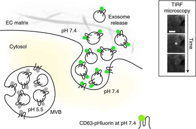
Exosomes are small extracellular vesicles with a diameter of 40-150 nm, and are implicated in cellular homeostasis and cell-cell communication. They can be secreted in bulk in response to cell-extrinsic and cell-intrinsic signals that cause multivesicular body (MVB) fusion with the plasma membrane (PM). However, research on the regulation of exosome release is hampered by the failure of current methods to capture the dynamics of exosome release. Here we describe how live imaging with tetraspanin-based pH-sensitive fluorescent reporters can quantify the MVB-PM fusion rate of single cells. Our approach enables identification of exogenous stimuli, signaling pathways, and fusion complexes, and can map subcellular sites of fusion events. In addition, dual-color imaging can be used to assess simultaneous release of different cargo by MVB exocytosis. This protocol describes the complete imaging experiment, consisting of transient expression of tetraspanin reporters (2 d), live-cell (dual-color) total internal reflection fluorescence microscopy (30-60 min per condition), and semiautomatic image analysis by using a newly developed ImageJ macro (±30 min per condition).
中文翻译:

多囊体质膜融合的实时成像,以量化外泌体从单细胞释放。
外泌体是直径为40-150 nm的小细胞外囊泡,与细胞稳态和细胞-细胞通讯有关。它们可响应细胞外和细胞内信号大量分泌,从而引起多囊体(MVB)与质膜(PM)融合。然而,目前方法无法捕获外泌体释放的动力学,阻碍了外泌体释放调控的研究。在这里,我们描述了如何使用基于四跨膜蛋白的pH敏感的荧光报道分子进行实时成像,以量化单个细胞的MVB-PM融合率。我们的方法能够识别外源性刺激,信号传导途径和融合复合物,并可以绘制融合事件的亚细胞位点。此外,双色成像可用于评估MVB胞吐作用同时释放不同货物。
更新日期:2019-12-17
中文翻译:
多囊体质膜融合的实时成像,以量化外泌体从单细胞释放。
外泌体是直径为40-150 nm的小细胞外囊泡,与细胞稳态和细胞-细胞通讯有关。它们可响应细胞外和细胞内信号大量分泌,从而引起多囊体(MVB)与质膜(PM)融合。然而,目前方法无法捕获外泌体释放的动力学,阻碍了外泌体释放调控的研究。在这里,我们描述了如何使用基于四跨膜蛋白的pH敏感的荧光报道分子进行实时成像,以量化单个细胞的MVB-PM融合率。我们的方法能够识别外源性刺激,信号传导途径和融合复合物,并可以绘制融合事件的亚细胞位点。此外,双色成像可用于评估MVB胞吐作用同时释放不同货物。


 京公网安备 11010802027423号