当前位置:
X-MOL 学术
›
ACS Photonics
›
论文详情
Our official English website, www.x-mol.net, welcomes your
feedback! (Note: you will need to create a separate account there.)
Single-Photon, Time-Gated, Phasor-Based Fluorescence Lifetime Imaging through Highly Scattering Medium
ACS Photonics ( IF 6.5 ) Pub Date : 2019-12-27 , DOI: 10.1021/acsphotonics.9b00874 Rinat Ankri 1 , Arkaprabha Basu 1 , Arin Can Ulku 2 , Claudio Bruschini 2 , Edoardo Charbon 2 , Shimon Weiss 1 , Xavier Michalet 1
ACS Photonics ( IF 6.5 ) Pub Date : 2019-12-27 , DOI: 10.1021/acsphotonics.9b00874 Rinat Ankri 1 , Arkaprabha Basu 1 , Arin Can Ulku 2 , Claudio Bruschini 2 , Edoardo Charbon 2 , Shimon Weiss 1 , Xavier Michalet 1
Affiliation
|
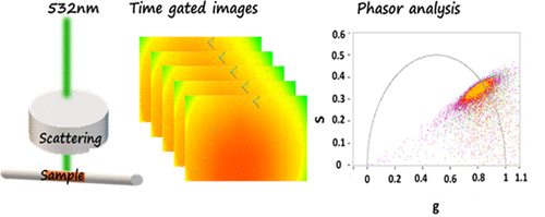
Fluorescence lifetime imaging (FLI) is increasingly recognized as a powerful tool for biochemical and cellular investigations, including in vivo applications. Fluorescence lifetime is an intrinsic characteristic of any fluorescent dye which, to a large extent, does not depend on excitation intensity and signal level. In particular, it allows distinguishing dyes with similar emission spectra, offering additional multiplexing capabilities. However, in vivo FLI in the visible range is complicated by the contamination by (i) tissue autofluorescence, which decreases contrast, and by (ii) light scattering and absorption in tissues, which significantly reduce fluorescence intensity and modify the temporal profile of the signal. Here, we demonstrate how these issues can be accounted for and overcome, using a new time-gated single-photon avalanche diode array camera, SwissSPAD2, combined with phasor analysis to provide a simple and fast visual method for lifetime imaging. In particular, we show how phasor dispersion increases with increasing scattering and/or decreasing fluorescence intensity. Next, we show that as long as the fluorescence signal of interest is larger than the phantom autofluorescence, the presence of a distinct lifetime can be clearly identified with appropriate background correction. We use these results to demonstrate the detection of A459 cells expressing the fluorescent protein mCyRFP1 through highly scattering and autofluorescent phantom layers. These results showcase the possibility to perform FLI in challenging conditions, using standard, bright, visible fluorophore or fluorescence proteins.
中文翻译:

通过高散射介质进行单光子、时间门控、基于相量的荧光寿命成像
荧光寿命成像 (FLI) 越来越被认为是生化和细胞研究(包括体内应用)的强大工具。荧光寿命是任何荧光染料的固有特性,它在很大程度上不依赖于激发强度和信号水平。特别是,它允许区分具有相似发射光谱的染料,提供额外的复用能力。然而,在体内可见范围内的 FLI 因 (i) 组织自发荧光(降低对比度)和 (ii) 组织中的光散射和吸收(显着降低荧光强度并改变信号的时间分布)的污染而变得复杂。在这里,我们展示了如何解决和克服这些问题,使用新的时间门控单光子雪崩二极管阵列相机 SwissSPAD2,结合相量分析,为寿命成像提供一种简单快速的视觉方法。特别是,我们展示了相量色散如何随着散射的增加和/或荧光强度的降低而增加。接下来,我们表明,只要感兴趣的荧光信号大于幻影自发荧光,就可以通过适当的背景校正清楚地识别出不同寿命的存在。我们使用这些结果来证明通过高度散射和自发荧光幻影层检测表达荧光蛋白 mCyRFP1 的 A459 细胞。这些结果展示了在具有挑战性的条件下使用标准、明亮、可见的荧光团或荧光蛋白进行 FLI 的可能性。
更新日期:2019-12-29
中文翻译:
通过高散射介质进行单光子、时间门控、基于相量的荧光寿命成像
荧光寿命成像 (FLI) 越来越被认为是生化和细胞研究(包括体内应用)的强大工具。荧光寿命是任何荧光染料的固有特性,它在很大程度上不依赖于激发强度和信号水平。特别是,它允许区分具有相似发射光谱的染料,提供额外的复用能力。然而,在体内可见范围内的 FLI 因 (i) 组织自发荧光(降低对比度)和 (ii) 组织中的光散射和吸收(显着降低荧光强度并改变信号的时间分布)的污染而变得复杂。在这里,我们展示了如何解决和克服这些问题,使用新的时间门控单光子雪崩二极管阵列相机 SwissSPAD2,结合相量分析,为寿命成像提供一种简单快速的视觉方法。特别是,我们展示了相量色散如何随着散射的增加和/或荧光强度的降低而增加。接下来,我们表明,只要感兴趣的荧光信号大于幻影自发荧光,就可以通过适当的背景校正清楚地识别出不同寿命的存在。我们使用这些结果来证明通过高度散射和自发荧光幻影层检测表达荧光蛋白 mCyRFP1 的 A459 细胞。这些结果展示了在具有挑战性的条件下使用标准、明亮、可见的荧光团或荧光蛋白进行 FLI 的可能性。


 京公网安备 11010802027423号