当前位置:
X-MOL 学术
›
ACS Biomater. Sci. Eng.
›
论文详情
Our official English website, www.x-mol.net, welcomes your
feedback! (Note: you will need to create a separate account there.)
Micropatterned Poly(ethylene glycol) Islands Disrupt Endothelial Cell-Substrate Interactions Differently from Microporous Membranes.
ACS Biomaterials Science & Engineering ( IF 5.4 ) Pub Date : 2020-01-03 , DOI: 10.1021/acsbiomaterials.9b01584 Zahra Allahyari 1, 2 , Shayan Gholizadeh 1, 2 , Henry H Chung 2 , Luis F Delgadillo 3 , Thomas R Gaborski 1, 2, 3
ACS Biomaterials Science & Engineering ( IF 5.4 ) Pub Date : 2020-01-03 , DOI: 10.1021/acsbiomaterials.9b01584 Zahra Allahyari 1, 2 , Shayan Gholizadeh 1, 2 , Henry H Chung 2 , Luis F Delgadillo 3 , Thomas R Gaborski 1, 2, 3
Affiliation

|
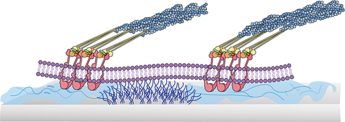
Porous membranes are ubiquitous in cell coculture and tissue-on-a-chip studies. These materials are predominantly chosen for their semipermeable and size exclusion properties to restrict or permit transmigration and cell–cell communication. However, previous studies have shown that pore size, spacing, and orientation affect cell behavior including extracellular matrix production and migration. The mechanism behind this behavior is not fully understood. In this study, we fabricated micropatterned nonfouling poly(ethylene glycol) (PEG) islands to mimic pore openings to decouple the effect of surface discontinuity from potential grip on the vertical contact area provided by pore wall edges. Similar to previous findings on porous membranes, we found that the PEG islands hindered fibronectin fibrillogenesis with cells on patterned substrates producing shorter fibrils. Additionally, cell migration speed over micropatterned PEG islands was greater than unpatterned controls, suggesting that disruption of cell–substrate interactions by PEG islands promoted a more dynamic and migratory behavior, similar to enhanced cell migration on microporous membranes. Preferred cellular directionality during migration was nearly indistinguishable between substrates with identically patterned PEG islands and previously reported behavior over micropores of the same geometry, further confirming disruption of cell–substrate interactions as a common mechanism behind the cellular responses on these substrates. Interestingly, compared to respective controls, there were differences in cell spreading and a lower increase in migration speed over PEG islands compared to prior results on micropores with identical feature size and spacing. This suggests that membrane pores not only disrupt cell–substrate interactions but also provide additional physical factors that affect cellular response.
中文翻译:

微图案化的聚(乙二醇)岛不同于微孔膜,破坏了内皮细胞-底物的相互作用。
多孔膜在细胞共培养和芯片上组织研究中无处不在。选择这些材料主要是因为它们具有半透性和尺寸排阻特性,以限制或允许迁移和细胞间通讯。但是,以前的研究表明,孔径,间距和方向会影响细胞行为,包括细胞外基质的产生和迁移。此行为背后的机制尚未完全了解。在这项研究中,我们制造了微图案的防污聚乙二醇(PEG)岛,以模仿孔的开口,从而将表面不连续性的影响与孔壁边缘提供的垂直接触区域上的潜在附着力分离开来。与先前在多孔膜上的发现相似,我们发现,PEG岛阻碍了纤连蛋白的原纤维形成,其中图案化底物上的细胞产生了较短的原纤维。此外,微图案化PEG岛上的细胞迁移速度要比未图案化的对照大,这表明PEG岛对细胞-底物相互作用的破坏促进了更动态和迁移的行为,类似于微孔膜上增强的细胞迁移。在迁移过程中,具有相同图案的PEG岛的底物与先前报道的具有相同几何形状的微孔之间的行为几乎无法区分。在这些底物上,细胞与底物相互作用的破坏是这些底物上细胞反应背后的共同机制,这进一步证实了细胞-底物相互作用的破坏。有趣的是,与各个控件相比,与先前在具有相同特征尺寸和间距的微孔上获得的结果相比,在PEG岛上存在细胞扩散差异和迁移速度增加较低的问题。这表明膜孔不仅破坏了细胞与底物的相互作用,而且还提供了影响细胞反应的其他物理因素。
更新日期:2020-01-04
中文翻译:

微图案化的聚(乙二醇)岛不同于微孔膜,破坏了内皮细胞-底物的相互作用。
多孔膜在细胞共培养和芯片上组织研究中无处不在。选择这些材料主要是因为它们具有半透性和尺寸排阻特性,以限制或允许迁移和细胞间通讯。但是,以前的研究表明,孔径,间距和方向会影响细胞行为,包括细胞外基质的产生和迁移。此行为背后的机制尚未完全了解。在这项研究中,我们制造了微图案的防污聚乙二醇(PEG)岛,以模仿孔的开口,从而将表面不连续性的影响与孔壁边缘提供的垂直接触区域上的潜在附着力分离开来。与先前在多孔膜上的发现相似,我们发现,PEG岛阻碍了纤连蛋白的原纤维形成,其中图案化底物上的细胞产生了较短的原纤维。此外,微图案化PEG岛上的细胞迁移速度要比未图案化的对照大,这表明PEG岛对细胞-底物相互作用的破坏促进了更动态和迁移的行为,类似于微孔膜上增强的细胞迁移。在迁移过程中,具有相同图案的PEG岛的底物与先前报道的具有相同几何形状的微孔之间的行为几乎无法区分。在这些底物上,细胞与底物相互作用的破坏是这些底物上细胞反应背后的共同机制,这进一步证实了细胞-底物相互作用的破坏。有趣的是,与各个控件相比,与先前在具有相同特征尺寸和间距的微孔上获得的结果相比,在PEG岛上存在细胞扩散差异和迁移速度增加较低的问题。这表明膜孔不仅破坏了细胞与底物的相互作用,而且还提供了影响细胞反应的其他物理因素。


 京公网安备 11010802027423号