当前位置:
X-MOL 学术
›
J. Biomed. Mater. Res. Part A
›
论文详情
Our official English website, www.x-mol.net, welcomes your
feedback! (Note: you will need to create a separate account there.)
Crayfish hemocyanin on chitin bone substitute scaffolds promotes the proliferation and osteogenic differentiation of human mesenchymal stem cells.
Journal of Biomedical Materials Research Part A ( IF 3.9 ) Pub Date : 2019-12-08 , DOI: 10.1002/jbm.a.36849 Benjamin Kruppke 1 , Jana Farack 1 , Simy Weil 2 , Eliahu David Aflalo 2, 3 , Dagmar Poláková 4 , Amir Sagi 2, 5 , Thomas Hanke 1
Journal of Biomedical Materials Research Part A ( IF 3.9 ) Pub Date : 2019-12-08 , DOI: 10.1002/jbm.a.36849 Benjamin Kruppke 1 , Jana Farack 1 , Simy Weil 2 , Eliahu David Aflalo 2, 3 , Dagmar Poláková 4 , Amir Sagi 2, 5 , Thomas Hanke 1
Affiliation
|
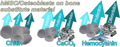
Crustacean chitin–hemocyanin–calcium mineral complexes were designed as bone biomimetics, with emphasis on their ability to bind or release calcium ions. Chitin scaffolds were prepared by dissolving chitin flakes in LiCl/dimethylacetamide, followed by gel formation and freeze‐drying. Some of these scaffolds were modified by incorporation of CaCO3. In some of the chitin–CaCO3 scaffolds, macroporosity was introduced by HCl treatment. Hemocyanin from the crayfish Cherax quadricarinatus was used to further modify the chitin scaffolds by dip coating. Cytocompatibility, cellular adherence and proliferation of human mesenchymal stem cells (hMSCs) were evaluated in terms of cell number as reflected in lactate dehydrogenase activity. The chitin, chitin–CaCO3, and porous chitin–CaCO3 scaffolds were all found to facilitate cell attachment. Hemocyanin dip‐coating of these scaffolds led to increased initial cell adhesion, enhanced proliferation, and osteogenic differentiation. Since the hemocyanin loading of the scaffolds was impaired by sterilization by gamma‐irradiation (as required for biomedical applications), the hemocyanin loading was performed on previously sterilized scaffolds. All scaffolds facilitated osteogenic differentiation of osteoblasts, with the highest cell ALP‐activity being found on hemocyanin‐modified porous chitin–CaCO3 scaffolds. Thus, chitin–hemocyanin scaffolds enhanced the initial stages of bone cell development and could serve as promising biomaterials for bone regeneration.
中文翻译:

甲壳素骨替代支架上的小龙虾血蓝蛋白促进人间充质干细胞的增殖和成骨分化。
甲壳类甲壳素-血蓝蛋白-钙矿物质复合物被设计为骨骼仿生物,重点是它们结合或释放钙离子的能力。通过将几丁质薄片溶解在氯化锂/二甲基乙酰胺中,然后形成凝胶和冷冻干燥来制备几丁质支架。这些支架中的一些通过加入 CaCO 3进行了修改。在一些几丁质-CaCO 3支架中,大孔隙是通过 HCl 处理引入的。来自小龙虾Cherax quadricarinatus 的血蓝蛋白用于通过浸涂进一步修饰几丁质支架。根据乳酸脱氢酶活性所反映的细胞数量评估人间充质干细胞 (hMSC) 的细胞相容性、细胞粘附和增殖。几丁质,几丁质-CaCO3和多孔几丁质-CaCO 3支架都被发现促进细胞附着。这些支架的血蓝蛋白浸涂导致初始细胞粘附增加、增殖增强和成骨分化。由于支架的血蓝蛋白负载受到伽马辐射灭菌(如生物医学应用所需)的影响,因此血蓝蛋白负载是在先前灭菌的支架上进行的。所有支架都促进了成骨细胞的成骨分化,在血蓝蛋白修饰的多孔甲壳素-CaCO 3支架上发现了最高的细胞 ALP 活性。因此,几丁质-血蓝蛋白支架增强了骨细胞发育的初始阶段,可以作为有希望的骨再生生物材料。
更新日期:2019-12-08
中文翻译:
甲壳素骨替代支架上的小龙虾血蓝蛋白促进人间充质干细胞的增殖和成骨分化。
甲壳类甲壳素-血蓝蛋白-钙矿物质复合物被设计为骨骼仿生物,重点是它们结合或释放钙离子的能力。通过将几丁质薄片溶解在氯化锂/二甲基乙酰胺中,然后形成凝胶和冷冻干燥来制备几丁质支架。这些支架中的一些通过加入 CaCO 3进行了修改。在一些几丁质-CaCO 3支架中,大孔隙是通过 HCl 处理引入的。来自小龙虾Cherax quadricarinatus 的血蓝蛋白用于通过浸涂进一步修饰几丁质支架。根据乳酸脱氢酶活性所反映的细胞数量评估人间充质干细胞 (hMSC) 的细胞相容性、细胞粘附和增殖。几丁质,几丁质-CaCO3和多孔几丁质-CaCO 3支架都被发现促进细胞附着。这些支架的血蓝蛋白浸涂导致初始细胞粘附增加、增殖增强和成骨分化。由于支架的血蓝蛋白负载受到伽马辐射灭菌(如生物医学应用所需)的影响,因此血蓝蛋白负载是在先前灭菌的支架上进行的。所有支架都促进了成骨细胞的成骨分化,在血蓝蛋白修饰的多孔甲壳素-CaCO 3支架上发现了最高的细胞 ALP 活性。因此,几丁质-血蓝蛋白支架增强了骨细胞发育的初始阶段,可以作为有希望的骨再生生物材料。


 京公网安备 11010802027423号