Our official English website, www.x-mol.net, welcomes your
feedback! (Note: you will need to create a separate account there.)
Morpho-molecular ex vivo detection and grading of non-muscle-invasive bladder cancer using forward imaging probe based multimodal optical coherence tomography and Raman spectroscopy.
Analyst ( IF 3.6 ) Pub Date : 2019-12-23 , DOI: 10.1039/c9an01911a Fabian Placzek 1 , Eliana Cordero Bautista , Simon Kretschmer , Lara M Wurster , Florian Knorr , Gerardo González-Cerdas , Mikael T Erkkilä , Patrick Stein , Çağlar Ataman , Gregers G Hermann , Karin Mogensen , Thomas Hasselager , Peter E Andersen , Hans Zappe , Jürgen Popp , Wolfgang Drexler , Rainer A Leitgeb , Iwan W Schie
Analyst ( IF 3.6 ) Pub Date : 2019-12-23 , DOI: 10.1039/c9an01911a Fabian Placzek 1 , Eliana Cordero Bautista , Simon Kretschmer , Lara M Wurster , Florian Knorr , Gerardo González-Cerdas , Mikael T Erkkilä , Patrick Stein , Çağlar Ataman , Gregers G Hermann , Karin Mogensen , Thomas Hasselager , Peter E Andersen , Hans Zappe , Jürgen Popp , Wolfgang Drexler , Rainer A Leitgeb , Iwan W Schie
Affiliation

|
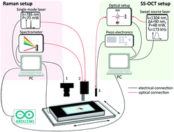
Non-muscle-invasive bladder cancer affects millions of people worldwide, resulting in significant discomfort to the patient and potential death. Today, cystoscopy is the gold standard for bladder cancer assessment, using white light endoscopy to detect tumor suspected lesion areas, followed by resection of these areas and subsequent histopathological evaluation. Not only does the pathological examination take days, but due to the invasive nature, the performed biopsy can result in significant harm to the patient. Nowadays, optical modalities, such as optical coherence tomography (OCT) and Raman spectroscopy (RS), have proven to detect cancer in real time and can provide more detailed clinical information of a lesion, e.g. its penetration depth (stage) and the differentiation of the cells (grade). In this paper, we present an ex vivo study performed with a combined piezoelectric tube-based OCT-probe and fiber optic RS-probe imaging system that allows large field-of-view imaging of bladder biopsies, using both modalities and co-registered visualization, detection and grading of cancerous bladder lesions. In the present study, 119 examined biopsies were characterized, showing that fiber-optic based OCT provides a sensitivity of 78% and a specificity of 69% for the detection of non-muscle-invasive bladder cancer, while RS, on the other hand, provides a sensitivity of 81% and a specificity of 61% for the grading of low- and high-grade tissues. Moreover, the study shows that a piezoelectric tube-based OCT probe can have significant endurance, suitable for future long-lasting in vivo applications. These results also indicate that combined OCT and RS fiber probe-based characterization offers an exciting possibility for label-free and morpho-chemical optical biopsies for bladder cancer diagnostics.
中文翻译:

使用正向成像探针的多模态光学相干断层扫描和拉曼光谱技术对非肌肉浸润性膀胱癌进行形态分子离体检测和分级。
非肌肉浸润性膀胱癌影响全世界数百万人,导致患者明显不适并可能导致死亡。如今,膀胱镜检查已成为评估膀胱癌的金标准,它使用白光内窥镜检查来检测可疑的肿瘤病变区域,然后切除这些区域并随后进行组织病理学评估。病理检查不仅要花费数天,而且由于其侵入性,所进行的活检可能会对患者造成重大伤害。如今,光学模式(如光学相干断层扫描(OCT)和拉曼光谱(RS))已被证明可以实时检测癌症,并且可以提供病变的更详细的临床信息,例如病变的穿透深度(阶段)和肿瘤的分化。单元格(等级)。在本文中,我们介绍了结合基于压电管的OCT探头和光纤RS探头成像系统进行的离体研究,该方法可使用模式和共同注册的可视化,检测和分级功能,对膀胱活检进行大视野成像癌性病变。在本研究中,对119项检查过的活检进行了表征,表明基于光纤的OCT对于检测非肌肉浸润性膀胱癌提供了78%的灵敏度和69%的特异性,而RS另一方面,对低度和高级组织的分级提供了81%的灵敏度和61%的特异性。此外,研究表明,基于压电管的OCT探针具有显着的耐用性,适用于未来的长期体内应用。
更新日期:2020-02-17
中文翻译:

使用正向成像探针的多模态光学相干断层扫描和拉曼光谱技术对非肌肉浸润性膀胱癌进行形态分子离体检测和分级。
非肌肉浸润性膀胱癌影响全世界数百万人,导致患者明显不适并可能导致死亡。如今,膀胱镜检查已成为评估膀胱癌的金标准,它使用白光内窥镜检查来检测可疑的肿瘤病变区域,然后切除这些区域并随后进行组织病理学评估。病理检查不仅要花费数天,而且由于其侵入性,所进行的活检可能会对患者造成重大伤害。如今,光学模式(如光学相干断层扫描(OCT)和拉曼光谱(RS))已被证明可以实时检测癌症,并且可以提供病变的更详细的临床信息,例如病变的穿透深度(阶段)和肿瘤的分化。单元格(等级)。在本文中,我们介绍了结合基于压电管的OCT探头和光纤RS探头成像系统进行的离体研究,该方法可使用模式和共同注册的可视化,检测和分级功能,对膀胱活检进行大视野成像癌性病变。在本研究中,对119项检查过的活检进行了表征,表明基于光纤的OCT对于检测非肌肉浸润性膀胱癌提供了78%的灵敏度和69%的特异性,而RS另一方面,对低度和高级组织的分级提供了81%的灵敏度和61%的特异性。此外,研究表明,基于压电管的OCT探针具有显着的耐用性,适用于未来的长期体内应用。


 京公网安备 11010802027423号