Our official English website, www.x-mol.net, welcomes your
feedback! (Note: you will need to create a separate account there.)
Sensitive Multicolor Visual Detection of Exosomes via Dual Signal Amplification Strategy of Enzyme-Catalyzed Metallization of Au Nanorods and Hybridization Chain Reaction.
ACS Sensors ( IF 8.2 ) Pub Date : 2019-12-17 , DOI: 10.1021/acssensors.9b01644 Yingzhi Zhang 1 , Danni Wang 1 , Shuai Yue 2 , Yanbing Lu 2 , Chunguang Yang 1 , Jin Fang 2 , Zhangrun Xu 1
ACS Sensors ( IF 8.2 ) Pub Date : 2019-12-17 , DOI: 10.1021/acssensors.9b01644 Yingzhi Zhang 1 , Danni Wang 1 , Shuai Yue 2 , Yanbing Lu 2 , Chunguang Yang 1 , Jin Fang 2 , Zhangrun Xu 1
Affiliation

|
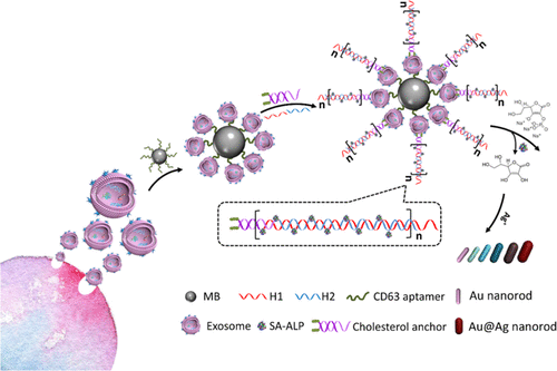
Exosomes as nanosized vesicles have been recognized as potential noninvasive biomarkers for early cancer diagnosis. Herein, we presented a sensitive multicolor visual method for exosome detection based on enzyme-induced silver deposition on gold nanorods (Au NRs). To achieve highly sensitive determination of exosomes, hybridization chain reaction (HCR) was employed to introduce more alkaline phosphatase (ALP) for signal amplification. First, exosomes were captured by magnetic bead-labeled CD63 aptamer, and, then, cholesterol-modified DNA probes were spontaneously inserted into the exosomal lipid membrane. The ends of the DNA probes act as the initiator to trigger the HCR for signal amplification. Finally, with the help of HCR, increased sites led to enhanced ALP loading and thus boosted the ascorbic acid generation. Silver ions were reduced by ascorbic acid, and silver shells were formed on Au NRs, giving rise to the blue shift of the longitudinal localized surface plasmon resonance peak. Correspondingly, the concentration of exosomes can be obviously distinguished with naked eyes via the vivid color variation. Due to the dual signal amplification of HCR and metallization of Au NRs, highly sensitive detection for exosomes were realized with detection limits as low as 1.6 × 10² particles/μL by UV-vis spectroscopy and 9 × 10³ particles/μL by naked eyes. Compared to the reported colorimetric methods for exosome quantification, visualization based on plentiful color tonalities is the most captivating merit of our approach, and HCR-induced signal amplification highlights the virtue of the strategy. The applicability of the method was validated by the analysis of clinical samples.
中文翻译:

酶催化金纳米棒金属化和杂交链反应的双信号放大策略,用于外来体的灵敏多色视觉检测。
外来体作为纳米大小的囊泡已被认为是用于早期癌症诊断的潜在非侵入性生物标志物。在这里,我们提出了一种基于酶诱导的金纳米棒(Au NRs)上的银沉积的外来体检测的灵敏多色视觉方法。为了实现高灵敏度的外泌体测定,采用了杂交链反应(HCR)引入更多的碱性磷酸酶(ALP)进行信号放大。首先,用磁珠标记的CD63适体捕获外泌体,然后将胆固醇修饰的DNA探针自发插入外泌体脂质膜。DNA探针的末端充当引发HCR进行信号放大的引发剂。最后,在HCR的帮助下,增加的位点导致ALP负载增加,从而促进了抗坏血酸的产生。抗坏血酸还原了银离子,并且在Au NRs上形成了银壳,从而引起了纵向局部表面等离振子共振峰的蓝移。相应地,通过生动的颜色变化可以用肉眼明显区分外泌体的浓度。由于HCR的双重信号放大和Au NRs的金属化,实现了对外泌体的高灵敏度检测,紫外可见光谱检测限低至1.6×10²颗粒/μL,肉眼检测限低至9×103颗粒/μL。与报告的比色法定量的外来体相比,基于丰富的色彩色调的可视化是我们方法最吸引人的优点,而HCR诱导的信号放大突出了该策略的优点。
更新日期:2019-12-18
中文翻译:

酶催化金纳米棒金属化和杂交链反应的双信号放大策略,用于外来体的灵敏多色视觉检测。
外来体作为纳米大小的囊泡已被认为是用于早期癌症诊断的潜在非侵入性生物标志物。在这里,我们提出了一种基于酶诱导的金纳米棒(Au NRs)上的银沉积的外来体检测的灵敏多色视觉方法。为了实现高灵敏度的外泌体测定,采用了杂交链反应(HCR)引入更多的碱性磷酸酶(ALP)进行信号放大。首先,用磁珠标记的CD63适体捕获外泌体,然后将胆固醇修饰的DNA探针自发插入外泌体脂质膜。DNA探针的末端充当引发HCR进行信号放大的引发剂。最后,在HCR的帮助下,增加的位点导致ALP负载增加,从而促进了抗坏血酸的产生。抗坏血酸还原了银离子,并且在Au NRs上形成了银壳,从而引起了纵向局部表面等离振子共振峰的蓝移。相应地,通过生动的颜色变化可以用肉眼明显区分外泌体的浓度。由于HCR的双重信号放大和Au NRs的金属化,实现了对外泌体的高灵敏度检测,紫外可见光谱检测限低至1.6×10²颗粒/μL,肉眼检测限低至9×103颗粒/μL。与报告的比色法定量的外来体相比,基于丰富的色彩色调的可视化是我们方法最吸引人的优点,而HCR诱导的信号放大突出了该策略的优点。


 京公网安备 11010802027423号