当前位置:
X-MOL 学术
›
Mol. Pharmaceutics
›
论文详情
Our official English website, www.x-mol.net, welcomes your
feedback! (Note: you will need to create a separate account there.)
Targeting Endothelin Receptors in a Murine Model of Myocardial Infarction Using a Small Molecular Fluorescent Probe.
Molecular Pharmaceutics ( IF 4.5 ) Pub Date : 2019-12-18 , DOI: 10.1021/acs.molpharmaceut.9b00810 Melanie A Kimm 1 , Helena Haas 1 , Miriam Stölting 2 , Michael Kuhlmann 3 , Christiane Geyer 2 , Sarah Glasl 4 , Michael Schäfers 3 , Vasilis Ntziachristos 4 , Moritz Wildgruber 1, 2 , Carsten Höltke 2
Molecular Pharmaceutics ( IF 4.5 ) Pub Date : 2019-12-18 , DOI: 10.1021/acs.molpharmaceut.9b00810 Melanie A Kimm 1 , Helena Haas 1 , Miriam Stölting 2 , Michael Kuhlmann 3 , Christiane Geyer 2 , Sarah Glasl 4 , Michael Schäfers 3 , Vasilis Ntziachristos 4 , Moritz Wildgruber 1, 2 , Carsten Höltke 2
Affiliation

|
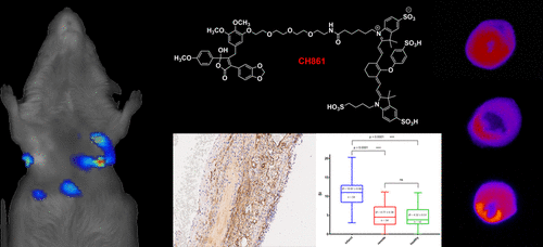
The endothelin (ET) axis plays a pivotal role in cardiovascular diseases. Enhanced levels of circulating ET-1 have been correlated with an inferior clinical outcome after myocardial infarction (MI) in humans. Thus, the evaluation of endothelin-A receptor (ETAR) expression over time in the course of myocardial injury and healing may offer valuable information toward the understanding of the ET axis involvement in MI. We developed an approach to track the expression of ETAR with a customized molecular imaging probe in a murine model of MI. The small molecular probe based on the ETAR-selective antagonist 3-(1,3-benzodioxol-5-yl)-5-hydroxy-5-(4-methoxyphenyl)-4-[(3,4,5-trimethoxyphenyl)methyl]-2(5H)-furanone (PD156707) was labeled with fluorescent dye, IRDye800cw. Mice undergoing permanent ligation of the left anterior descending artery (LAD) were investigated at day 1, 7, and 21 post surgery after receiving an intravenous injection of the ETAR probe. Cryosections of explanted hearts were analyzed by cryotome-based CCD, and fluorescence reflectance imaging (FRI) and fluorescence signal intensities (SI) were extracted. Fluorescence-mediated tomography (FMT) imaging was performed to visualize probe distribution in the target region in vivo. An enhanced fluorescence signal intensity in the infarct area was detected in cryoCCD images as early as day 1 after surgery and intensified up to 21 days post MI. FRI was capable of detecting significantly enhanced SI in infarcted regions of hearts 7 days after surgery. In vivo imaging by FMT localized enhanced SI in the apex region of infarcted mouse hearts. We verified the localization of the probe and ETAR within the infarct area by immunohistochemistry (IHC). In addition, neovascularized areas were found in the affected myocardium by CD31 staining. Our study demonstrates that the applied fluorescent probe is capable of delineating ETAR expression over time in affected murine myocardium after MI in vivo and ex vivo.
中文翻译:

使用小分子荧光探针在心肌梗塞的小鼠模型中靶向内皮素受体。
内皮素(ET)轴在心血管疾病中起关键作用。循环中ET-1水平的提高与人类心肌梗死(MI)后的不良临床预后相关。因此,评估心肌损伤和愈合过程中内皮素A受体(ETAR)的表达可能会为了解MI中的ET轴提供有价值的信息。我们开发了一种方法,可以使用定制的分子成像探针在MI小鼠模型中追踪ETAR的表达。基于ETAR选择性拮抗剂3-(1,3-苯并二恶唑-5-基)-5-羟基-5-(4-甲氧基苯基)-4-[(3,4,5-三甲氧基苯基)甲基的小分子探针用荧光染料IRDye800cw标记] -2(5H)-呋喃酮(PD156707)。在接受静脉内注射ETAR探针后,在术后第1、7和21天研究了永久性结扎左前降支动脉(LAD)的小鼠。通过基于冷冻切片机的CCD对离体心脏的冷冻切片进行分析,并提取荧光反射成像(FRI)和荧光信号强度(SI)。进行荧光介导的断层扫描(FMT)成像以可视化探针在体内靶区域中的分布。最早在手术后第1天,在cryoCCD图像中就检测到了梗死区域荧光信号强度的增强,并在MI后长达21天的时间里增强了。手术7天后,FRI能够检测到心脏梗死区域的SI明显增强。通过FMT的体内成像将梗死小鼠心脏的心尖区域中增强的SI定位。我们通过免疫组织化学(IHC)验证了梗塞区域内探针和ETAR的定位。另外,通过CD31染色在受影响的心肌中发现了新血管形成的区域。我们的研究表明,所应用的荧光探针能够在体内和离体心肌梗死后随时间描绘出ETAR在受影响的鼠心肌中的表达。
更新日期:2019-12-19
中文翻译:

使用小分子荧光探针在心肌梗塞的小鼠模型中靶向内皮素受体。
内皮素(ET)轴在心血管疾病中起关键作用。循环中ET-1水平的提高与人类心肌梗死(MI)后的不良临床预后相关。因此,评估心肌损伤和愈合过程中内皮素A受体(ETAR)的表达可能会为了解MI中的ET轴提供有价值的信息。我们开发了一种方法,可以使用定制的分子成像探针在MI小鼠模型中追踪ETAR的表达。基于ETAR选择性拮抗剂3-(1,3-苯并二恶唑-5-基)-5-羟基-5-(4-甲氧基苯基)-4-[(3,4,5-三甲氧基苯基)甲基的小分子探针用荧光染料IRDye800cw标记] -2(5H)-呋喃酮(PD156707)。在接受静脉内注射ETAR探针后,在术后第1、7和21天研究了永久性结扎左前降支动脉(LAD)的小鼠。通过基于冷冻切片机的CCD对离体心脏的冷冻切片进行分析,并提取荧光反射成像(FRI)和荧光信号强度(SI)。进行荧光介导的断层扫描(FMT)成像以可视化探针在体内靶区域中的分布。最早在手术后第1天,在cryoCCD图像中就检测到了梗死区域荧光信号强度的增强,并在MI后长达21天的时间里增强了。手术7天后,FRI能够检测到心脏梗死区域的SI明显增强。通过FMT的体内成像将梗死小鼠心脏的心尖区域中增强的SI定位。我们通过免疫组织化学(IHC)验证了梗塞区域内探针和ETAR的定位。另外,通过CD31染色在受影响的心肌中发现了新血管形成的区域。我们的研究表明,所应用的荧光探针能够在体内和离体心肌梗死后随时间描绘出ETAR在受影响的鼠心肌中的表达。


 京公网安备 11010802027423号