Our official English website, www.x-mol.net, welcomes your feedback! (Note: you will need to create a separate account there.)
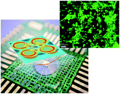
A microelectrode array chip for osteogenic differentiation of mesenchymal stem cells under electrical stimulation.
Lab on a Chip ( IF 6.1 ) Pub Date : 2019-12-18 , DOI: 10.1039/c9lc01081e Tianyang Zheng 1 , Zhizhong Zhang 1 , Rong Zhu 1 , Dong Sun 2
Lab on a Chip ( IF 6.1 ) Pub Date : 2019-12-18 , DOI: 10.1039/c9lc01081e Tianyang Zheng 1 , Zhizhong Zhang 1 , Rong Zhu 1 , Dong Sun 2
Affiliation
|
Electrical stimulation (ES) as an easy and effective inducing method has been widely used in induction differentiation of stem cells, e.g. osteogenic differentiation of mesenchymal stem cells (MSCs) for bone healing and bone tissue therapies. However, the micro-effect of an inhomogeneous electric field has rarely been investigated for ES in induction differentiation, and conventionally used ex situ assays may preclude accurate assessment due to variation from cell inoculation and treatments. Here, a novel electrical stimulation method with a microelectrode array chip is proposed for osteogenic differentiation of MSCs. The electric field applied onto the MSCs by the microelectrode array is designed similarly with a natural aggregation distribution of differentiated MSCs. The proposed ES method accelerates osteoblast proliferation and differentiation in the electrode array region and generates a larger amount of mineralized deposits, which are assayed via in situ alizarin red staining and morphology observation as well as immunocytochemistry. In addition, this method allows a direct in situ assessment to compare the osteogenic differentiation of MSCs with and without ES on a single chip to avoid culture environment difference. The method provides a fundamental platform for investigating induced differentiation of stem cells and allows integration with multifunctional cell assays to achieve in situ tracking for the differentiation process of stem cells.
中文翻译:

在电刺激下用于间充质干细胞成骨分化的微电极阵列芯片。
电刺激(ES)是一种简便有效的诱导方法,已广泛用于干细胞的诱导分化,例如间充质干细胞(MSCs)的成骨分化,用于骨愈合和骨组织治疗。然而,对于诱导分化中的ES,很少研究不均匀电场的微效应,并且由于细胞接种和处理方法的差异,常规使用的异位分析可能无法进行准确的评估。在此,提出了一种新的具有微电极阵列芯片的电刺激方法用于MSC的成骨分化。通过微电极阵列施加到MSC上的电场的设计与分化MSC的自然聚集分布相似。拟议的ES方法可加速成骨细胞在电极阵列区域的增殖和分化,并产生大量矿化沉积物,这些沉积物可通过原位茜素红染色和形态学观察以及免疫细胞化学进行测定。此外,这种方法可以直接进行原位评估,以比较在单个芯片上有无ES的MSC的成骨分化,从而避免培养环境的差异。该方法为研究干细胞的诱导分化提供了基础平台,并允许与多功能细胞测定法整合以实现对干细胞分化过程的原位追踪。通过原位茜素红染色和形态观察以及免疫细胞化学进行检测。此外,这种方法可以直接进行原位评估,以比较在单个芯片上有无ES的MSC的成骨分化,从而避免培养环境的差异。该方法为研究干细胞的诱导分化提供了基础平台,并允许与多功能细胞测定法整合以实现对干细胞分化过程的原位追踪。通过原位茜素红染色和形态观察以及免疫细胞化学进行检测。此外,这种方法可以直接进行原位评估,以比较在单个芯片上有无ES的MSC的成骨分化,从而避免培养环境的差异。该方法为研究干细胞的诱导分化提供了基础平台,并允许与多功能细胞测定法整合以实现对干细胞分化过程的原位追踪。
更新日期:2020-02-13
中文翻译:
在电刺激下用于间充质干细胞成骨分化的微电极阵列芯片。
电刺激(ES)是一种简便有效的诱导方法,已广泛用于干细胞的诱导分化,例如间充质干细胞(MSCs)的成骨分化,用于骨愈合和骨组织治疗。然而,对于诱导分化中的ES,很少研究不均匀电场的微效应,并且由于细胞接种和处理方法的差异,常规使用的异位分析可能无法进行准确的评估。在此,提出了一种新的具有微电极阵列芯片的电刺激方法用于MSC的成骨分化。通过微电极阵列施加到MSC上的电场的设计与分化MSC的自然聚集分布相似。拟议的ES方法可加速成骨细胞在电极阵列区域的增殖和分化,并产生大量矿化沉积物,这些沉积物可通过原位茜素红染色和形态学观察以及免疫细胞化学进行测定。此外,这种方法可以直接进行原位评估,以比较在单个芯片上有无ES的MSC的成骨分化,从而避免培养环境的差异。该方法为研究干细胞的诱导分化提供了基础平台,并允许与多功能细胞测定法整合以实现对干细胞分化过程的原位追踪。通过原位茜素红染色和形态观察以及免疫细胞化学进行检测。此外,这种方法可以直接进行原位评估,以比较在单个芯片上有无ES的MSC的成骨分化,从而避免培养环境的差异。该方法为研究干细胞的诱导分化提供了基础平台,并允许与多功能细胞测定法整合以实现对干细胞分化过程的原位追踪。通过原位茜素红染色和形态观察以及免疫细胞化学进行检测。此外,这种方法可以直接进行原位评估,以比较在单个芯片上有无ES的MSC的成骨分化,从而避免培养环境的差异。该方法为研究干细胞的诱导分化提供了基础平台,并允许与多功能细胞测定法整合以实现对干细胞分化过程的原位追踪。


 京公网安备 11010802027423号