当前位置:
X-MOL 学术
›
Dent. Mater.
›
论文详情
Our official English website, www.x-mol.net, welcomes your feedback! (Note: you will need to create a separate account there.)
Hydroxyapatite-based cements induce different apatite formation in radicular dentin.
Dental Materials ( IF 5 ) Pub Date : 2019-12-07 , DOI: 10.1016/j.dental.2019.11.023 Manuel Toledano-Osorio 1 , Fátima S Aguilera 1 , Raquel Osorio 1 , Esther Muñoz-Soto 1 , Mayra C Pérez-Álvarez 2 , Modesto T López-López 3 , Christopher D Lynch 4 , Manuel Toledano 1
Dental Materials ( IF 5 ) Pub Date : 2019-12-07 , DOI: 10.1016/j.dental.2019.11.023 Manuel Toledano-Osorio 1 , Fátima S Aguilera 1 , Raquel Osorio 1 , Esther Muñoz-Soto 1 , Mayra C Pérez-Álvarez 2 , Modesto T López-López 3 , Christopher D Lynch 4 , Manuel Toledano 1
Affiliation
|
OBJECTIVE
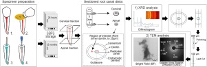
To investigate crystallinity and ultrastructure of the formed hydroxyapatite at radicular cervical and apical dentin after being treated with three different canal sealers.
METHODS
Cervical and apical root dentin surfaces were treated with two experimental hydroxyapatite-based sealers, containing sodium hydroxide (calcypatite) or zinc oxide (oxipatite) and an epoxy resin-based canal sealer (AH Plus); gutta-percha without sealer was included as control. Dentin surfaces were studied by X-ray diffraction and transmission electron microscopy through selected area diffraction and bright-field imaging after 24h and 12m of storage.
RESULTS
Root cervical dentin treated with calcypatite and oxipatite produced poor crystallinity of new minerals, wide amorphous phase and non-stoichiometry. Reflections at the 002 plane and the corresponding diffraction rings attained lower values in the Scherrer equation and the Scherrer-Wilson equation in samples treated with both HAp-based sealers than in specimens without sealer or with AH Plus. At root cervical dentin treated with calcypatite, shorter and wider crystallite size formations and lower crystals grain size were found, if compared to those encountered at oxipatite treated dentin. Oxipatite attained improved crystallographic atomic order and less structural variation in both distances and angles. Apical dentin treated with oxipatite attained preferred grain orientation with polycrystalline lattices.
SIGNIFICANCE
The immature crystallites formed in dentin treated with calcypatite and oxipatite will account for high hydroxyapatite solubility and remineralizing activity. New polycrystalline formations encountered in apical dentin treated with oxipatite may also produce high mechanical performance.
中文翻译:

羟基磷灰石基水泥在放射状牙本质中诱导不同的磷灰石形成。
目的研究经三种不同的管封闭剂处理后形成的羟基磷灰石在子宫颈和顶端牙本质的结晶度和超微结构。方法用两种基于羟基磷灰石的封闭剂处理宫颈和根尖牙本质表面,封闭剂中含有氢氧化钠(方解石)或氧化锌(氧磷钙石)和环氧树脂基牙本质封闭剂(AH Plus)。不含密封剂的牙胶加糖胶作为对照。储存24h和12m后,通过X射线衍射和透射电子显微镜通过选择区域衍射和明场成像研究牙本质表面。结果经钙磷灰石和氧磷灰石处理的根颈型牙本质产生了较差的新矿物质结晶,较宽的无定形相和非化学计量比。在使用两种基于HAp的密封剂处理的样品中,与没有密封剂或AH Plus的样品相比,在002平面上的反射和相应的衍射环在Scherrer方程和Scherrer-Wilson方程中获得的值更低。与用氧化磷钙石处理过的牙本质相比,在经钙磷灰石处理过的宫颈根部牙本质上发现了更短,更宽的微晶尺寸形成和更低的晶体晶粒尺寸。磷灰石获得了改进的晶体原子序,并且在距离和角度上的结构变化都较小。用氧磷灰石处理的根尖牙本质具有多晶格的优选晶粒取向。意义钙磷灰石和氧磷灰石处理过的牙本质中形成的未成熟微晶将说明羟基磷灰石具有较高的溶解度和再矿化活性。
更新日期:2019-12-07
中文翻译:
羟基磷灰石基水泥在放射状牙本质中诱导不同的磷灰石形成。
目的研究经三种不同的管封闭剂处理后形成的羟基磷灰石在子宫颈和顶端牙本质的结晶度和超微结构。方法用两种基于羟基磷灰石的封闭剂处理宫颈和根尖牙本质表面,封闭剂中含有氢氧化钠(方解石)或氧化锌(氧磷钙石)和环氧树脂基牙本质封闭剂(AH Plus)。不含密封剂的牙胶加糖胶作为对照。储存24h和12m后,通过X射线衍射和透射电子显微镜通过选择区域衍射和明场成像研究牙本质表面。结果经钙磷灰石和氧磷灰石处理的根颈型牙本质产生了较差的新矿物质结晶,较宽的无定形相和非化学计量比。在使用两种基于HAp的密封剂处理的样品中,与没有密封剂或AH Plus的样品相比,在002平面上的反射和相应的衍射环在Scherrer方程和Scherrer-Wilson方程中获得的值更低。与用氧化磷钙石处理过的牙本质相比,在经钙磷灰石处理过的宫颈根部牙本质上发现了更短,更宽的微晶尺寸形成和更低的晶体晶粒尺寸。磷灰石获得了改进的晶体原子序,并且在距离和角度上的结构变化都较小。用氧磷灰石处理的根尖牙本质具有多晶格的优选晶粒取向。意义钙磷灰石和氧磷灰石处理过的牙本质中形成的未成熟微晶将说明羟基磷灰石具有较高的溶解度和再矿化活性。


 京公网安备 11010802027423号