Our official English website, www.x-mol.net, welcomes your feedback! (Note: you will need to create a separate account there.)
A microfluidic device for both on-chip dialysis protein crystallization and in situ X-ray diffraction.
Lab on a Chip ( IF 6.1 ) Pub Date : 2019-12-05 , DOI: 10.1039/c9lc00651f Niels Junius 1 , Sofia Jaho 1 , Yoann Sallaz-Damaz 1 , Franck Borel 1 , Jean-Baptiste Salmon 2 , Monika Budayova-Spano 1
Lab on a Chip ( IF 6.1 ) Pub Date : 2019-12-05 , DOI: 10.1039/c9lc00651f Niels Junius 1 , Sofia Jaho 1 , Yoann Sallaz-Damaz 1 , Franck Borel 1 , Jean-Baptiste Salmon 2 , Monika Budayova-Spano 1
Affiliation

|
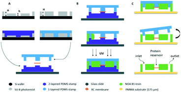
This paper reports a versatile microfluidic chip developed for on-chip crystallization of proteins through the dialysis method and in situ X-ray diffraction experiments. A microfabrication process enabling the integration of regenerated cellulose dialysis membranes between two layers of the microchip is thoroughly described. We also describe a rational approach for optimizing on-chip protein crystallization via chemical composition and temperature control, allowing the crystal size, number and quality to be tailored. Combining optically transparent microfluidics and dialysis provides both precise control over the experiment and reversible exploration of the crystallization conditions. In addition, the materials composing the microfluidic chip were tested for their transparency to X-rays in order to assess their compatibility for in situ diffraction data collection. Background scattering was evaluated using a synchrotron X-ray source and the background noise generated by our microfluidic device was compared to that produced by commercial crystallization plates used for diffraction experiments at room temperature. Once crystals of 3 model proteins (lysozyme, IspE, and insulin) were grown on-chip, the microchip was mounted onto the beamline and partial diffraction data sets were collected in situ from several isomorphous crystals and were merged to a complete data set for structure determination. We therefore propose a robust and inexpensive way to fabricate microchips that cover the whole pipeline from crystal growth to the beam and does not require any handling of the protein crystals prior to the diffraction experiment, allowing the collection of crystallographic data at room temperature for solving the three-dimensional structure of the proteins under study. The results presented here allow serial crystallography experiments on synchrotrons and X-ray lasers under dynamically controllable sample conditions to be observed using the developed microchips.
中文翻译:

用于芯片上透析蛋白结晶和原位X射线衍射的微流控设备。
本文报道了一种通用的微流控芯片,该芯片通过透析方法和原位X射线衍射实验开发用于蛋白质的芯片结晶。彻底描述了使再生纤维素透析膜在微芯片的两层之间集成的微加工过程。我们还描述了一种通过化学成分和温度控制来优化片上蛋白质结晶的合理方法,从而可以调整晶体的大小,数量和质量。将光学透明的微流控技术与渗析技术相结合,可提供对实验的精确控制以及对结晶条件的可逆探索。此外,测试组成微流体芯片的材料对X射线的透明性,以评估其与原位衍射数据收集的兼容性。使用同步加速器X射线源评估背景散射,并将我们的微流体装置产生的背景噪声与室温下用于衍射实验的商用结晶板产生的背景噪声进行比较。一旦3种模型蛋白(溶菌酶,IspE和胰岛素)的晶体在芯片上生长,将微芯片安装到光束线上,并从几个同构晶体中就地收集部分衍射数据集,并合并为完整的结构数据集决心。因此,我们提出了一种健壮而廉价的方法来制造微芯片,该芯片覆盖从晶体生长到光束的整个管道,并且不需要在衍射实验之前对蛋白质晶体进行任何处理,从而可以在室温下收集晶体学数据来解决蛋白质的三维结构。此处提供的结果允许使用开发的微芯片在动态可控的样品条件下在同步加速器和X射线激光器上进行串行晶体学实验。
更新日期:2020-02-13
中文翻译:

用于芯片上透析蛋白结晶和原位X射线衍射的微流控设备。
本文报道了一种通用的微流控芯片,该芯片通过透析方法和原位X射线衍射实验开发用于蛋白质的芯片结晶。彻底描述了使再生纤维素透析膜在微芯片的两层之间集成的微加工过程。我们还描述了一种通过化学成分和温度控制来优化片上蛋白质结晶的合理方法,从而可以调整晶体的大小,数量和质量。将光学透明的微流控技术与渗析技术相结合,可提供对实验的精确控制以及对结晶条件的可逆探索。此外,测试组成微流体芯片的材料对X射线的透明性,以评估其与原位衍射数据收集的兼容性。使用同步加速器X射线源评估背景散射,并将我们的微流体装置产生的背景噪声与室温下用于衍射实验的商用结晶板产生的背景噪声进行比较。一旦3种模型蛋白(溶菌酶,IspE和胰岛素)的晶体在芯片上生长,将微芯片安装到光束线上,并从几个同构晶体中就地收集部分衍射数据集,并合并为完整的结构数据集决心。因此,我们提出了一种健壮而廉价的方法来制造微芯片,该芯片覆盖从晶体生长到光束的整个管道,并且不需要在衍射实验之前对蛋白质晶体进行任何处理,从而可以在室温下收集晶体学数据来解决蛋白质的三维结构。此处提供的结果允许使用开发的微芯片在动态可控的样品条件下在同步加速器和X射线激光器上进行串行晶体学实验。


 京公网安备 11010802027423号