当前位置:
X-MOL 学术
›
J. Photochem. Photobiol. B Biol.
›
论文详情
Our official English website, www.x-mol.net, welcomes your
feedback! (Note: you will need to create a separate account there.)
Imaging mitochondria and plasma membrane in live cells using solvatochromic styrylpyridines.
Journal of Photochemistry and Photobiology B: Biology ( IF 3.9 ) Pub Date : 2019-12-05 , DOI: 10.1016/j.jphotobiol.2019.111732 Tarushyam Mukherjee 1 , M Aravintha Siva 2 , Komal Bajaj 1 , Virupakshi Soppina 2 , Sriram Kanvah 1
Journal of Photochemistry and Photobiology B: Biology ( IF 3.9 ) Pub Date : 2019-12-05 , DOI: 10.1016/j.jphotobiol.2019.111732 Tarushyam Mukherjee 1 , M Aravintha Siva 2 , Komal Bajaj 1 , Virupakshi Soppina 2 , Sriram Kanvah 1
Affiliation
|
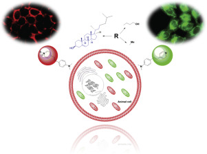
Investigating the dynamics of different biomolecules in the cellular milieu through microscopic imaging has gained paramount importance in the last decade. Continuous developments in the field of microscopy are paralleled by the design and synthesis of fluorophores that target specific compartments within a cell. In this study, we have synthesized four fluorescent styrene derivatives, a neutral styrylpridine, three cationic styrylpyridinium probes with and without cholesterol tether, and investigated their absorption, emission, and cellular imaging properties. The fluorophores show solvatochromic emission attributed to intramolecular charge transfer from donor to acceptor with an emission range of 500-600 nm. The fluorescent cholesterol conjugate labels plasma membrane effectively while the fluorophores devoid of the cholesterol tether label mitochondria. Cholesterol conjugate also shows strong interaction with liposome membrane. Furthermore, the fluorophores alsotrack the mitochondria in live cells with high specificity. Cell viability assay showed overall non-toxic nature of the probes even at higher fluorophore concentrations. Through sidearm modifications, keeping the fluorescent core intact, we successfully targeted specific subcellular compartments of neuronal (N2a) and non-neuronal (HeLa) mammalian cell lines. This strategy of using a single molecular scaffold with subtle substitutions could be ideal in generating a variety of fluorophores targeting other subcellular compartments.
中文翻译:

使用溶剂变色苯乙烯基吡啶对活细胞中的线粒体和质膜进行成像。
在过去的十年中,通过显微镜成像研究细胞环境中不同生物分子的动力学已变得至关重要。显微镜领域的不断发展与靶向细胞内特定区室的荧光团的设计和合成并行。在这项研究中,我们合成了四个荧光苯乙烯衍生物,一个中性苯乙烯基吡啶,三个带有和不带有胆固醇束缚带的阳离子苯乙烯基吡啶鎓探针,并研究了它们的吸收,发射和细胞成像特性。荧光团显示溶剂致变色发射,这归因于分子内电荷从供体到受体的分子转移,发射范围为500-600 nm。荧光胆固醇偶联物可有效标记质膜,而荧光团不含胆固醇束缚线粒体。胆固醇结合物也显示出与脂质体膜的强相互作用。此外,荧光团还以高特异性追踪活细胞中的线粒体。细胞活力分析表明,即使在较高的荧光团浓度下,探针的总体无毒性质。通过侧臂修饰,使荧光核心保持完整,我们成功地靶向了神经元(N2a)和非神经元(HeLa)哺乳动物细胞系的特定亚细胞区室。使用具有微妙取代的单个分子支架的这种策略对于产生靶向其他亚细胞区室的各种荧光团可能是理想的。胆固醇结合物也显示出与脂质体膜的强相互作用。此外,荧光团还以高特异性追踪活细胞中的线粒体。细胞活力分析表明,即使在较高的荧光团浓度下,探针的总体无毒性质。通过侧臂修饰,使荧光核心保持完整,我们成功地靶向了神经元(N2a)和非神经元(HeLa)哺乳动物细胞系的特定亚细胞区室。使用具有微妙取代的单个分子支架的这种策略对于产生靶向其他亚细胞区室的各种荧光团可能是理想的。胆固醇结合物也显示出与脂质体膜的强相互作用。此外,荧光团还以高特异性追踪活细胞中的线粒体。细胞活力分析表明,即使在较高的荧光团浓度下,探针的总体无毒性质。通过侧臂修饰,保持荧光核心完整,我们成功地靶向了神经元(N2a)和非神经元(HeLa)哺乳动物细胞系的特定亚细胞区室。使用具有微小取代的单个分子支架的这种策略对于产生靶向其他亚细胞区室的各种荧光团可能是理想的。细胞活力分析表明,即使在较高的荧光团浓度下,探针的总体无毒性质。通过侧臂修饰,使荧光核心保持完整,我们成功地靶向了神经元(N2a)和非神经元(HeLa)哺乳动物细胞系的特定亚细胞区室。使用具有微妙取代的单个分子支架的这种策略对于产生靶向其他亚细胞区室的各种荧光团可能是理想的。细胞活力分析表明,即使在较高的荧光团浓度下,探针的总体无毒性质。通过侧臂修饰,保持荧光核心完整,我们成功地靶向了神经元(N2a)和非神经元(HeLa)哺乳动物细胞系的特定亚细胞区室。使用具有微妙取代的单个分子支架的这种策略对于产生靶向其他亚细胞区室的各种荧光团可能是理想的。
更新日期:2019-12-05
中文翻译:
使用溶剂变色苯乙烯基吡啶对活细胞中的线粒体和质膜进行成像。
在过去的十年中,通过显微镜成像研究细胞环境中不同生物分子的动力学已变得至关重要。显微镜领域的不断发展与靶向细胞内特定区室的荧光团的设计和合成并行。在这项研究中,我们合成了四个荧光苯乙烯衍生物,一个中性苯乙烯基吡啶,三个带有和不带有胆固醇束缚带的阳离子苯乙烯基吡啶鎓探针,并研究了它们的吸收,发射和细胞成像特性。荧光团显示溶剂致变色发射,这归因于分子内电荷从供体到受体的分子转移,发射范围为500-600 nm。荧光胆固醇偶联物可有效标记质膜,而荧光团不含胆固醇束缚线粒体。胆固醇结合物也显示出与脂质体膜的强相互作用。此外,荧光团还以高特异性追踪活细胞中的线粒体。细胞活力分析表明,即使在较高的荧光团浓度下,探针的总体无毒性质。通过侧臂修饰,使荧光核心保持完整,我们成功地靶向了神经元(N2a)和非神经元(HeLa)哺乳动物细胞系的特定亚细胞区室。使用具有微妙取代的单个分子支架的这种策略对于产生靶向其他亚细胞区室的各种荧光团可能是理想的。胆固醇结合物也显示出与脂质体膜的强相互作用。此外,荧光团还以高特异性追踪活细胞中的线粒体。细胞活力分析表明,即使在较高的荧光团浓度下,探针的总体无毒性质。通过侧臂修饰,使荧光核心保持完整,我们成功地靶向了神经元(N2a)和非神经元(HeLa)哺乳动物细胞系的特定亚细胞区室。使用具有微妙取代的单个分子支架的这种策略对于产生靶向其他亚细胞区室的各种荧光团可能是理想的。胆固醇结合物也显示出与脂质体膜的强相互作用。此外,荧光团还以高特异性追踪活细胞中的线粒体。细胞活力分析表明,即使在较高的荧光团浓度下,探针的总体无毒性质。通过侧臂修饰,保持荧光核心完整,我们成功地靶向了神经元(N2a)和非神经元(HeLa)哺乳动物细胞系的特定亚细胞区室。使用具有微小取代的单个分子支架的这种策略对于产生靶向其他亚细胞区室的各种荧光团可能是理想的。细胞活力分析表明,即使在较高的荧光团浓度下,探针的总体无毒性质。通过侧臂修饰,使荧光核心保持完整,我们成功地靶向了神经元(N2a)和非神经元(HeLa)哺乳动物细胞系的特定亚细胞区室。使用具有微妙取代的单个分子支架的这种策略对于产生靶向其他亚细胞区室的各种荧光团可能是理想的。细胞活力分析表明,即使在较高的荧光团浓度下,探针的总体无毒性质。通过侧臂修饰,保持荧光核心完整,我们成功地靶向了神经元(N2a)和非神经元(HeLa)哺乳动物细胞系的特定亚细胞区室。使用具有微妙取代的单个分子支架的这种策略对于产生靶向其他亚细胞区室的各种荧光团可能是理想的。


 京公网安备 11010802027423号