Our official English website, www.x-mol.net, welcomes your
feedback! (Note: you will need to create a separate account there.)
Detection of collagens by multispectral optoacoustic tomography as an imaging biomarker for Duchenne muscular dystrophy.
Nature Medicine ( IF 58.7 ) Pub Date : 2019-12-02 , DOI: 10.1038/s41591-019-0669-y Adrian P Regensburger 1 , Lina M Fonteyne 2 , Jörg Jüngert 1 , Alexandra L Wagner 1 , Teresa Gerhalter 3, 4, 5 , Armin M Nagel 3, 4, 5 , Rafael Heiss 3 , Florian Flenkenthaler 2 , Matthias Qurashi 6 , Markus F Neurath 6, 7 , Nikolai Klymiuk 2 , Elisabeth Kemter 2 , Thomas Fröhlich 2 , Michael Uder 3 , Joachim Woelfle 1 , Wolfgang Rascher 1 , Regina Trollmann 1 , Eckhard Wolf 2 , Maximilian J Waldner 6, 8 , Ferdinand Knieling 1
Nature Medicine ( IF 58.7 ) Pub Date : 2019-12-02 , DOI: 10.1038/s41591-019-0669-y Adrian P Regensburger 1 , Lina M Fonteyne 2 , Jörg Jüngert 1 , Alexandra L Wagner 1 , Teresa Gerhalter 3, 4, 5 , Armin M Nagel 3, 4, 5 , Rafael Heiss 3 , Florian Flenkenthaler 2 , Matthias Qurashi 6 , Markus F Neurath 6, 7 , Nikolai Klymiuk 2 , Elisabeth Kemter 2 , Thomas Fröhlich 2 , Michael Uder 3 , Joachim Woelfle 1 , Wolfgang Rascher 1 , Regina Trollmann 1 , Eckhard Wolf 2 , Maximilian J Waldner 6, 8 , Ferdinand Knieling 1
Affiliation
|
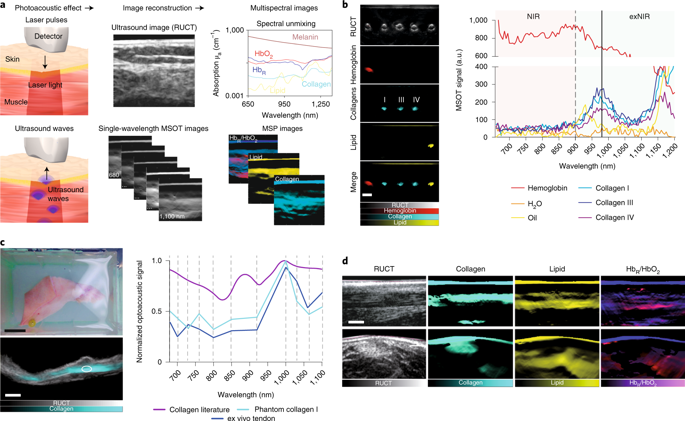
Biomarkers for monitoring of disease progression and response to therapy are lacking for muscle diseases such as Duchenne muscular dystrophy. Noninvasive in vivo molecular imaging with multispectral optoacoustic tomography (MSOT) uses pulsed laser light to induce acoustic pressure waves, enabling the visualization of endogenous chromophores. Here we describe an application of MSOT, in which illumination in the near- and extended near-infrared ranges from 680-1,100 nm enables the visualization and quantification of collagen content. We first demonstrated the feasibility of this approach to noninvasive quantification of tissue fibrosis in longitudinal studies in a large-animal Duchenne muscular dystrophy model in pigs, and then applied this approach to pediatric patients. MSOT-derived collagen content measurements in skeletal muscle were highly correlated to the functional status of the patients and provided additional information on molecular features as compared to magnetic resonance imaging. This study highlights the potential of MSOT imaging as a noninvasive, age-independent biomarker for the implementation and monitoring of newly developed therapies in muscular diseases.
中文翻译:

通过多光谱光声断层扫描检测胶原蛋白作为杜氏肌营养不良症的成像生物标志物。
杜氏肌营养不良等肌肉疾病缺乏用于监测疾病进展和治疗反应的生物标志物。具有多光谱光声断层扫描 (MSOT) 的无创体内分子成像使用脉冲激光诱导声压波,从而实现内源生色团的可视化。在这里,我们描述了 MSOT 的应用,其中在 680-1,100 nm 的近红外和扩展近红外范围内的照明使胶原蛋白含量的可视化和量化成为可能。我们首先在猪的大型动物杜氏肌营养不良模型的纵向研究中证明了这种方法对组织纤维化进行无创量化的可行性,然后将这种方法应用于儿科患者。与磁共振成像相比,骨骼肌中 MSOT 衍生的胶原蛋白含量测量与患者的功能状态高度相关,并提供了有关分子特征的额外信息。本研究强调了 MSOT 成像作为一种非侵入性、与年龄无关的生物标志物的潜力,可用于实施和监测新开发的肌肉疾病疗法。
更新日期:2019-12-02
中文翻译:
通过多光谱光声断层扫描检测胶原蛋白作为杜氏肌营养不良症的成像生物标志物。
杜氏肌营养不良等肌肉疾病缺乏用于监测疾病进展和治疗反应的生物标志物。具有多光谱光声断层扫描 (MSOT) 的无创体内分子成像使用脉冲激光诱导声压波,从而实现内源生色团的可视化。在这里,我们描述了 MSOT 的应用,其中在 680-1,100 nm 的近红外和扩展近红外范围内的照明使胶原蛋白含量的可视化和量化成为可能。我们首先在猪的大型动物杜氏肌营养不良模型的纵向研究中证明了这种方法对组织纤维化进行无创量化的可行性,然后将这种方法应用于儿科患者。与磁共振成像相比,骨骼肌中 MSOT 衍生的胶原蛋白含量测量与患者的功能状态高度相关,并提供了有关分子特征的额外信息。本研究强调了 MSOT 成像作为一种非侵入性、与年龄无关的生物标志物的潜力,可用于实施和监测新开发的肌肉疾病疗法。


 京公网安备 11010802027423号