Our official English website, www.x-mol.net, welcomes your
feedback! (Note: you will need to create a separate account there.)
Three-dimensional spatially resolved geometrical and functional models of human liver tissue reveal new aspects of NAFLD progression.
Nature Medicine ( IF 58.7 ) Pub Date : 2019-12-02 , DOI: 10.1038/s41591-019-0660-7 Fabián Segovia-Miranda 1 , Hernán Morales-Navarrete 1 , Michael Kücken 2 , Vincent Moser 3 , Sarah Seifert 1 , Urska Repnik 1 , Fabian Rost 2, 4 , Mario Brosch 3, 5 , Alexander Hendricks 6 , Sebastian Hinz 6 , Christoph Röcken 7 , Dieter Lütjohann 8 , Yannis Kalaidzidis 1, 9 , Clemens Schafmayer 6 , Lutz Brusch 2 , Jochen Hampe 3, 5 , Marino Zerial 1
Nature Medicine ( IF 58.7 ) Pub Date : 2019-12-02 , DOI: 10.1038/s41591-019-0660-7 Fabián Segovia-Miranda 1 , Hernán Morales-Navarrete 1 , Michael Kücken 2 , Vincent Moser 3 , Sarah Seifert 1 , Urska Repnik 1 , Fabian Rost 2, 4 , Mario Brosch 3, 5 , Alexander Hendricks 6 , Sebastian Hinz 6 , Christoph Röcken 7 , Dieter Lütjohann 8 , Yannis Kalaidzidis 1, 9 , Clemens Schafmayer 6 , Lutz Brusch 2 , Jochen Hampe 3, 5 , Marino Zerial 1
Affiliation
|
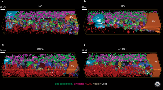
Early disease diagnosis is key to the effective treatment of diseases. Histopathological analysis of human biopsies is the gold standard to diagnose tissue alterations. However, this approach has low resolution and overlooks 3D (three-dimensional) structural changes resulting from functional alterations. Here, we applied multiphoton imaging, 3D digital reconstructions and computational simulations to generate spatially resolved geometrical and functional models of human liver tissue at different stages of non-alcoholic fatty liver disease (NAFLD). We identified a set of morphometric cellular and tissue parameters correlated with disease progression, and discover profound topological defects in the 3D bile canalicular (BC) network. Personalized biliary fluid dynamic simulations predicted an increased pericentral biliary pressure and micro-cholestasis, consistent with elevated cholestatic biomarkers in patients' sera. Our spatially resolved models of human liver tissue can contribute to high-definition medicine by identifying quantitative multiparametric cellular and tissue signatures to define disease progression and provide new insights into NAFLD pathophysiology.
中文翻译:

人体肝脏组织的三维空间分辨几何和功能模型揭示了 NAFLD 进展的新方面。
疾病的早期诊断是有效治疗疾病的关键。人体活检组织病理学分析是诊断组织改变的金标准。然而,这种方法具有低分辨率并且忽略了由功能改变导致的 3D(三维)结构变化。在这里,我们应用多光子成像、3D 数字重建和计算模拟来生成非酒精性脂肪性肝病 (NAFLD) 不同阶段的人体肝脏组织的空间分辨几何和功能模型。我们确定了一组与疾病进展相关的形态计量细胞和组织参数,并在 3D 胆小管 (BC) 网络中发现了深刻的拓扑缺陷。个性化的胆汁流体动力学模拟预测中心周围胆汁压力和微胆汁淤积增加,这与患者血清中胆汁淤积生物标志物升高一致。我们的人类肝组织空间分辨模型可以通过识别定量的多参数细胞和组织特征来定义疾病进展并为 NAFLD 病理生理学提供新的见解,从而为高清医学做出贡献。
更新日期:2019-12-02
中文翻译:
人体肝脏组织的三维空间分辨几何和功能模型揭示了 NAFLD 进展的新方面。
疾病的早期诊断是有效治疗疾病的关键。人体活检组织病理学分析是诊断组织改变的金标准。然而,这种方法具有低分辨率并且忽略了由功能改变导致的 3D(三维)结构变化。在这里,我们应用多光子成像、3D 数字重建和计算模拟来生成非酒精性脂肪性肝病 (NAFLD) 不同阶段的人体肝脏组织的空间分辨几何和功能模型。我们确定了一组与疾病进展相关的形态计量细胞和组织参数,并在 3D 胆小管 (BC) 网络中发现了深刻的拓扑缺陷。个性化的胆汁流体动力学模拟预测中心周围胆汁压力和微胆汁淤积增加,这与患者血清中胆汁淤积生物标志物升高一致。我们的人类肝组织空间分辨模型可以通过识别定量的多参数细胞和组织特征来定义疾病进展并为 NAFLD 病理生理学提供新的见解,从而为高清医学做出贡献。


 京公网安备 11010802027423号