当前位置:
X-MOL 学术
›
Microsyst. Nanoeng.
›
论文详情
Our official English website, www.x-mol.net, welcomes your
feedback! (Note: you will need to create a separate account there.)
Quantitative phase imaging of erythrocytes under microfluidic constriction in a high refractive index medium reveals water content changes.
Microsystems & Nanoengineering ( IF 7.3 ) Pub Date : 2019-12-02 , DOI: 10.1038/s41378-019-0113-y Han Sang Park 1 , Will J Eldridge 1 , Wen-Hsuan Yang 2, 3, 4 , Michael Crose 1 , Silvia Ceballos 1 , John D Roback 5 , Jen-Tsan Ashley Chi 2, 3 , Adam Wax 1
Microsystems & Nanoengineering ( IF 7.3 ) Pub Date : 2019-12-02 , DOI: 10.1038/s41378-019-0113-y Han Sang Park 1 , Will J Eldridge 1 , Wen-Hsuan Yang 2, 3, 4 , Michael Crose 1 , Silvia Ceballos 1 , John D Roback 5 , Jen-Tsan Ashley Chi 2, 3 , Adam Wax 1
Affiliation
|
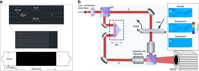
Changes in the deformability of red blood cells can reveal a range of pathologies. For example, cells which have been stored for transfusion are known to exhibit progressively impaired deformability. Thus, this aspect of red blood cells has been characterized previously using a range of techniques. In this paper, we show a novel approach for examining the biophysical response of the cells with quantitative phase imaging. Specifically, optical volume changes are observed as the cells transit restrictive channels of a microfluidic chip in a high refractive index medium. The optical volume changes indicate an increase of cell's internal density, ostensibly due to water displacement. Here, we characterize these changes over time for red blood cells from two subjects. By storage day 29, a significant decrease in the magnitude of optical volume change in response to mechanical stress was witnessed. The exchange of water with the environment due to mechanical stress is seen to modulate with storage time, suggesting a potential means for studying cell storage.
中文翻译:

在高折射率介质中微流体收缩下红细胞的定量相位成像揭示了水含量的变化。
红细胞变形能力的变化可以揭示一系列病理。例如,已知为输血而储存的细胞表现出逐渐受损的变形能力。因此,之前已经使用一系列技术表征了红细胞的这一方面。在本文中,我们展示了一种通过定量相位成像检查细胞生物物理反应的新方法。具体而言,当细胞在高折射率介质中通过微流控芯片的限制性通道时,观察到光学体积变化。光学体积变化表明细胞内部密度增加,表面上是由于水置换。在这里,我们表征了来自两个受试者的红细胞随时间的这些变化。到储存第 29 天,目睹了响应机械应力的光学体积变化幅度的显着下降。由于机械应力与环境的水交换被认为随储存时间而调节,这表明研究细胞储存的潜在手段。
更新日期:2019-12-02
中文翻译:
在高折射率介质中微流体收缩下红细胞的定量相位成像揭示了水含量的变化。
红细胞变形能力的变化可以揭示一系列病理。例如,已知为输血而储存的细胞表现出逐渐受损的变形能力。因此,之前已经使用一系列技术表征了红细胞的这一方面。在本文中,我们展示了一种通过定量相位成像检查细胞生物物理反应的新方法。具体而言,当细胞在高折射率介质中通过微流控芯片的限制性通道时,观察到光学体积变化。光学体积变化表明细胞内部密度增加,表面上是由于水置换。在这里,我们表征了来自两个受试者的红细胞随时间的这些变化。到储存第 29 天,目睹了响应机械应力的光学体积变化幅度的显着下降。由于机械应力与环境的水交换被认为随储存时间而调节,这表明研究细胞储存的潜在手段。


 京公网安备 11010802027423号