Our official English website, www.x-mol.net, welcomes your
feedback! (Note: you will need to create a separate account there.)
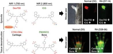
Fluorometric Imaging for Early Diagnosis and Prognosis of Rheumatoid Arthritis.
Advanced Science ( IF 14.3 ) Pub Date : 2019-12-01 , DOI: 10.1002/advs.201902267 Jeong Heon Lee 1 , Sang Youn Jung 2 , G Kate Park 1 , Kai Bao 1 , Hoon Hyun 3 , Georges El Fakhri 1 , Hak Soo Choi 1
Advanced Science ( IF 14.3 ) Pub Date : 2019-12-01 , DOI: 10.1002/advs.201902267 Jeong Heon Lee 1 , Sang Youn Jung 2 , G Kate Park 1 , Kai Bao 1 , Hoon Hyun 3 , Georges El Fakhri 1 , Hak Soo Choi 1
Affiliation
|
Early diagnosis and monitoring of disease progress are of significant importance in the effective treatment of rheumatoid arthritis (RA), because the continuing inflammation can lead to irreversible joint damage and systemic complications. However, applying imaging modalities for the prognosis of RA remains challenging, because no tissue-specific guidelines are available to monitor the progressive course of RA. In this study, fluorometric imaging of RA is reported using bioengineered targeted agents of the blood vessel, bone, and cartilage in combination with the customized optical fluorescence imaging system. Separate but simultaneous tissue-specific images of synovitis, cartilage destruction, and bone resorption are obtained from a mouse model of RA, which allows quantification of the prognosis of diseases at each stage. Thus, the fluorometric imaging of RA by using tissue-specific contrast agents plays a key role in the systemic treatment of RA by monitoring structural damage and disease progression.
中文翻译:

荧光成像用于类风湿性关节炎的早期诊断和预后。
早期诊断和监测疾病进展对于类风湿性关节炎(RA)的有效治疗具有重要意义,因为持续的炎症可导致不可逆的关节损伤和全身并发症。然而,应用影像学方法来预测 RA 的预后仍然具有挑战性,因为没有组织特异性指南可用于监测 RA 的进展过程。在这项研究中,报告了使用血管、骨骼和软骨的生物工程靶向药物结合定制的光学荧光成像系统进行 RA 荧光成像。从 RA 小鼠模型中获得滑膜炎、软骨破坏和骨吸收的单独但同时的组织特异性图像,从而可以量化每个阶段的疾病预后。因此,使用组织特异性造影剂对 RA 进行荧光成像,通过监测结构损伤和疾病进展,在 RA 的全身治疗中发挥着关键作用。
更新日期:2019-12-02
中文翻译:
荧光成像用于类风湿性关节炎的早期诊断和预后。
早期诊断和监测疾病进展对于类风湿性关节炎(RA)的有效治疗具有重要意义,因为持续的炎症可导致不可逆的关节损伤和全身并发症。然而,应用影像学方法来预测 RA 的预后仍然具有挑战性,因为没有组织特异性指南可用于监测 RA 的进展过程。在这项研究中,报告了使用血管、骨骼和软骨的生物工程靶向药物结合定制的光学荧光成像系统进行 RA 荧光成像。从 RA 小鼠模型中获得滑膜炎、软骨破坏和骨吸收的单独但同时的组织特异性图像,从而可以量化每个阶段的疾病预后。因此,使用组织特异性造影剂对 RA 进行荧光成像,通过监测结构损伤和疾病进展,在 RA 的全身治疗中发挥着关键作用。


 京公网安备 11010802027423号