当前位置:
X-MOL 学术
›
Angew. Chem. Int. Ed.
›
论文详情
Our official English website, www.x-mol.net, welcomes your
feedback! (Note: you will need to create a separate account there.)
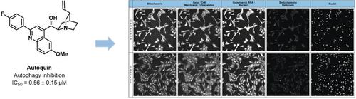
Image-Based Morphological Profiling Identifies a Lysosomotropic, Iron-Sequestering Autophagy Inhibitor.
Angewandte Chemie International Edition ( IF 16.1 ) Pub Date : 2019-11-26 , DOI: 10.1002/anie.201913712 Luca Laraia 1, 2 , Guillaume Garivet 1, 3 , Daniel J Foley 1, 4 , Nadine Kaiser 1, 3 , Sebastian Müller 5 , Sarah Zinken 1, 3 , Thomas Pinkert 6 , Julian Wilke 1, 3 , Dale Corkery 7 , Axel Pahl 8 , Sonja Sievers 8 , Petra Janning 1, 3 , Christoph Arenz 6 , Yaowen Wu 7 , Raphaël Rodriguez 5 , Herbert Waldmann 1, 3
Angewandte Chemie International Edition ( IF 16.1 ) Pub Date : 2019-11-26 , DOI: 10.1002/anie.201913712 Luca Laraia 1, 2 , Guillaume Garivet 1, 3 , Daniel J Foley 1, 4 , Nadine Kaiser 1, 3 , Sebastian Müller 5 , Sarah Zinken 1, 3 , Thomas Pinkert 6 , Julian Wilke 1, 3 , Dale Corkery 7 , Axel Pahl 8 , Sonja Sievers 8 , Petra Janning 1, 3 , Christoph Arenz 6 , Yaowen Wu 7 , Raphaël Rodriguez 5 , Herbert Waldmann 1, 3
Affiliation
|
Chemical proteomics is widely applied in small-molecule target identification. However, in general it does not identify non-protein small-molecule targets, and thus, alternative methods for target identification are in high demand. We report the discovery of the autophagy inhibitor autoquin and the identification of its molecular mode of action using image-based morphological profiling in the cell painting assay. A compound-induced fingerprint representing changes in 579 cellular parameters revealed that autoquin accumulates in lysosomes and inhibits their fusion with autophagosomes. In addition, autoquin sequesters Fe2+ in lysosomes, resulting in an increase of lysosomal reactive oxygen species and ultimately cell death. Such a mechanism of action would have been challenging to unravel by current methods. This work demonstrates the potential of the cell painting assay to deconvolute modes of action of small molecules, warranting wider application in chemical biology.
中文翻译:

基于图像的形态分析可识别溶同质,铁螯合自噬抑制剂。
化学蛋白质组学被广泛应用于小分子目标识别中。然而,通常它不能鉴定非蛋白质的小分子靶标,因此,对靶标鉴定的替代方法有很高的要求。我们报告自噬抑制剂autoquin的发现,并在细胞绘画测定中使用基于图像的形态学分析对其分子作用方式进行鉴定。代表579个细胞参数变化的化合物诱导的指纹显示,自体喹啉在溶酶体中积累,并抑制了它们与自噬体的融合。此外,自动喹啉螯合溶酶体中的Fe2 +,导致溶酶体活性氧的增加,并最终导致细胞死亡。用当前的方法来解开这种作用机制将是具有挑战性的。
更新日期:2020-01-24
中文翻译:
基于图像的形态分析可识别溶同质,铁螯合自噬抑制剂。
化学蛋白质组学被广泛应用于小分子目标识别中。然而,通常它不能鉴定非蛋白质的小分子靶标,因此,对靶标鉴定的替代方法有很高的要求。我们报告自噬抑制剂autoquin的发现,并在细胞绘画测定中使用基于图像的形态学分析对其分子作用方式进行鉴定。代表579个细胞参数变化的化合物诱导的指纹显示,自体喹啉在溶酶体中积累,并抑制了它们与自噬体的融合。此外,自动喹啉螯合溶酶体中的Fe2 +,导致溶酶体活性氧的增加,并最终导致细胞死亡。用当前的方法来解开这种作用机制将是具有挑战性的。


 京公网安备 11010802027423号