当前位置:
X-MOL 学术
›
Biomater. Sci.
›
论文详情
Our official English website, www.x-mol.net, welcomes your
feedback! (Note: you will need to create a separate account there.)
3D biofabrication of microfiber-laden minispheroids: a facile 3D cell co-culturing system.
Biomaterials Science ( IF 5.8 ) Pub Date : 2019-11-25 , DOI: 10.1039/c9bm01189g Mingjun Xie 1 , Qing Gao , Jingjiang Qiu , Jianzhong Fu , Zichen Chen , Yong He
Biomaterials Science ( IF 5.8 ) Pub Date : 2019-11-25 , DOI: 10.1039/c9bm01189g Mingjun Xie 1 , Qing Gao , Jingjiang Qiu , Jianzhong Fu , Zichen Chen , Yong He
Affiliation
|
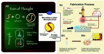
Hierarchical tissues composed of spheroid and fiber structures such as tumors, embryos and glomeruli widely exist in organisms. Methods have been developed to build spheroid and fiber structures, independently, as tissue models in vitro. However, it is still a challenge to print them simultaneously and integrated for effectively mimicking the complicated situations in vivo. Here, we propose a novel 3D cell co-culturing system, “microfiber-laden minispheroid”, applying two fluidic phenomena, namely the “rope coiling effect” and “electrohydrodynamics”, with a co-axial bioprinting nozzle and high voltage system. Gelatin methacryloyl (GelMA) hydrogels were extruded from the outer nozzle and separated by electrical attraction to form spheroids (∼1800 μm). GelMA mixed with sodium alginate was extruded from the inner nozzle to form fibers (∼180 μm) inside spheroids, whose morphology could be controlled by the ratio of inner and outer nozzle extruding flow rates. We analyzed the fabrication process and the material system in detail, verifying the fabrication feasibility and suitable microenvironment. The encapsulated cells possessed high viabilities. Importantly, the actin of human umbilical vein endothelial cells tended to elongate towards the co-cultured tumor cells, in contrast to the HUVECs cultured alone. We believe that “microfiber-laden minispheroids” could be a potential system for 3D cell co-culturing research in the future.
中文翻译:

载有超细纤维的微型球的3D生物制造:便捷的3D细胞共培养系统。
由球体和纤维结构组成的分层组织,例如肿瘤,胚胎和肾小球广泛存在于生物体中。已经开发了独立地构建球状和纤维结构的方法,作为体外组织模型。然而,同时打印它们并进行集成以有效地模拟体内的复杂情况仍然是一个挑战。。在这里,我们提出了一种新颖的3D细胞共培养系统,“微纤维负载的小球体”,通过同轴的生物打印喷嘴和高压系统,应用了两种流体现象,即“绳索卷曲效应”和“电流体动力学”。从外部喷嘴中挤出明胶甲基丙烯酰基(GelMA)水凝胶,并通过电引力将其分离,以形成球体(〜1800μm)。将与海藻酸钠混合的GelMA从内部喷嘴中挤出,以在球体内部形成纤维(〜180μm),其形态可通过内部和外部喷嘴挤出流量的比值来控制。我们详细分析了制造工艺和材料系统,验证了制造可行性和合适的微环境。包封的细胞具有高生存力。重要的,与单独培养的HUVEC相比,人脐静脉内皮细胞的肌动蛋白倾向于向共培养的肿瘤细胞延伸。我们相信,“超细纤维微型球体”将来可能成为3D细胞共培养研究的潜在系统。
更新日期:2019-11-25
中文翻译:
载有超细纤维的微型球的3D生物制造:便捷的3D细胞共培养系统。
由球体和纤维结构组成的分层组织,例如肿瘤,胚胎和肾小球广泛存在于生物体中。已经开发了独立地构建球状和纤维结构的方法,作为体外组织模型。然而,同时打印它们并进行集成以有效地模拟体内的复杂情况仍然是一个挑战。。在这里,我们提出了一种新颖的3D细胞共培养系统,“微纤维负载的小球体”,通过同轴的生物打印喷嘴和高压系统,应用了两种流体现象,即“绳索卷曲效应”和“电流体动力学”。从外部喷嘴中挤出明胶甲基丙烯酰基(GelMA)水凝胶,并通过电引力将其分离,以形成球体(〜1800μm)。将与海藻酸钠混合的GelMA从内部喷嘴中挤出,以在球体内部形成纤维(〜180μm),其形态可通过内部和外部喷嘴挤出流量的比值来控制。我们详细分析了制造工艺和材料系统,验证了制造可行性和合适的微环境。包封的细胞具有高生存力。重要的,与单独培养的HUVEC相比,人脐静脉内皮细胞的肌动蛋白倾向于向共培养的肿瘤细胞延伸。我们相信,“超细纤维微型球体”将来可能成为3D细胞共培养研究的潜在系统。


 京公网安备 11010802027423号